Physical Diagnosis PDX
Library
Cardiology Atlas Tutorial
Neurology Atlas Tutorial
Rheumatology Atlas Tutorial
Endocrinology Atlas Tutorial
Gastroenterology Atlas Tutorial
Pulmonology Atlas Tutorial
Infectious Disease Atlas Tutorial
Hematology-Oncology Atlas Tutorial
Nephrology Atlas Tutorial
Nails Atlas Tutorial
Hereditary Atlas Tutorial
Miscellaneous Atlas Tutorial
Cases Lectures Quizzes About
Library

Cardiology

Atlas Tutorial

Neurology

Atlas Tutorial

Rheumatology

Atlas Tutorial

Endocrinology

Atlas Tutorial

Gastroenterology

Atlas Tutorial

Pulmonology

Atlas Tutorial

Infectious Disease

Atlas Tutorial

Hematology-Oncology

Atlas Tutorial

Nephrology

Atlas Tutorial

Nails

Atlas Tutorial

Hereditary

Atlas Tutorial

Miscellaneous

Atlas Tutorial
Cases Lectures Quizzes About
  1. Home
  2. Cases
  3. Cirrhosis with a murmur

ccirrhosis5.png

File is not under priv/media.
Case 33 Gastroenterology

Cirrhosis with a murmur

Work it one clue at a time. The diagnosis stays hidden until you reveal it.

Clue 1

A middle-aged woman with a year-long history of Laennec’s cirrhosis (EtOH-related) comes under your care.  

Sure enough she has signs of liver disease on exam.

Clue 2

What else do you notice in the above video? The EJ is engorged. This is an unexpected finding in a patient with cirrhosis, where you would expect to see extravascular hypervolemia (eg, pitting edema, ascites) but not intravascular hypervolemia. Let’s take a closer look.

Clue 3

Intravascular volume is markedly elevated.  

Let’s take a listen to the heart: (best to use headphones or good computer speakers.)

Clue 4

What you’re hearing is a booming gallop. Our patient is so tachycardic it’s tough to distinguish between an S3 and an S4. However, the phonocardiogram can help.

Clue 5

Our patient has an S4 gallop.

Let’s look back at ascitic fluid results since the diagnosis of Laennec’s cirrhosis was made. SAAG >1.1, consistent with a portal hypertensive process.

Clue 6

But not all cases of portal hypertension are caused by cirrhosis. What do you think about the total protein (TP) concentration of the ascitic fluid?

Clue 7

Ascitic fluid total protein is greater than expected for classic cirrhosis, where there is a problem manufacturing protein. Ascitic TP in cirrhotic patients is usually <2.5 g/dL. Is there a condition that is associated with portal hypertension and ascites with TP >2.5?

Clue 8

One way to distinguish between cirrhosis and heart failure is to measure the hepatic venous pressure gradient (HVPG):

Clue 9

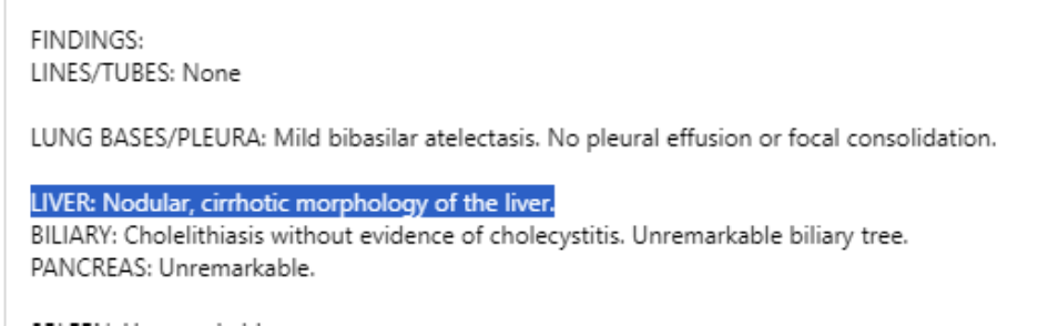
But with exam findings as clear as our patient’s elevated neck veins and the booming gallop, I think we have what we need to say the heart is driving this picture. But how do we explain the findings of cirrhosis on CT imaging?

Clue 10

This patient likely has “cardiac cirrhosis,” which occurs as a result of chronic congestion in the liver from elevated right-sided filling pressures. (The histology of cardiac cirrhosis is unique compared to other forms of cirrhosis).

End of case.

Teaching physical diagnosis through a library of real clinical findings.

  • About
  • Resources
  • Privacy
  • Acknowledgments