Physical Diagnosis PDX
Library
Cardiology Atlas Tutorial
Neurology Atlas Tutorial
Rheumatology Atlas Tutorial
Endocrinology Atlas Tutorial
Gastroenterology Atlas Tutorial
Pulmonology Atlas Tutorial
Infectious Disease Atlas Tutorial
Hematology-Oncology Atlas Tutorial
Nephrology Atlas Tutorial
Nails Atlas Tutorial
Hereditary Atlas Tutorial
Miscellaneous Atlas Tutorial
Cases Lectures Quizzes About
Library

Cardiology

Atlas Tutorial

Neurology

Atlas Tutorial

Rheumatology

Atlas Tutorial

Endocrinology

Atlas Tutorial

Gastroenterology

Atlas Tutorial

Pulmonology

Atlas Tutorial

Infectious Disease

Atlas Tutorial

Hematology-Oncology

Atlas Tutorial

Nephrology

Atlas Tutorial

Nails

Atlas Tutorial

Hereditary

Atlas Tutorial

Miscellaneous

Atlas Tutorial
Cases Lectures Quizzes About
  1. Home
  2. Cases
  3. Elevated JVP

Picture8-2.png

File is not under priv/media.
Case 27 Cardiology

Elevated JVP

Work it one clue at a time. The diagnosis stays hidden until you reveal it.

Clue 1

I was in the hospital with a group of students when a cardiologist spotted us. “Go see the patient in such and such room, you might like his exam.” 

So we go in blind.  

We notice that he’s young (30s). Next, we examine the neck. Here he is in the upright position.

Clue 2

JVP is elevated and Kussmaul’s sign is present. Normally the JVP goes down with inspiration.

In addition to Kussmaul’s sign, there is a second qualitative finding, better seen in this video. What part of the waveform catches your eye? Outward or inward?

Clue 3

The students start to name the things that could cause Kussmaul’s sign as well as giant a-waves. The as-yet unknown diagnosis is among their hypotheses. Before we listen, we anticipate. The ears can’t hear what the mind doesn’t know.

Clue 4

Over the apex there is a split S1. 

Next we hear a murmur over the left lower sternal border. Our collection of hypotheses allowed us to predict that this murmur might be present.

Clue 5

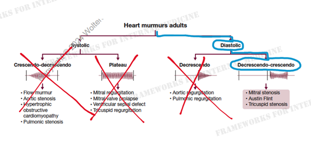
When determining the cause of a murmur, timing is key. Is it systolic or diastolic? This will cut our differential diagnosis in half.

Clue 6

Timing with the cardiac cycle allows us to determine that the murmur is occurring in diastole. It is also quite long in duration, which can serve as nice clue to a diastolic murmur (since diastole is longer than systole).

Clue 7

The next question we want to know is the shape of the murmur. Is it the classic decrescendo murmur of aortic regurgitation or does it get louder at the end? 

Our patient’s murmur clearly crescendos at the end of diastole.

Clue 8

So are we dealing with mitral or tricuspid stenosis? How can we tell? 

Did you appreciate that the murmur increases in intensity during inspiration? Carvallo’s sign tells us the lesion is on the right-side of the heart. Listen again.

Reveal next

Teaching physical diagnosis through a library of real clinical findings.

  • About
  • Resources
  • Privacy
  • Acknowledgments