Physical Diagnosis PDX
Library
Cardiology Atlas Tutorial
Neurology Atlas Tutorial
Rheumatology Atlas Tutorial
Endocrinology Atlas Tutorial
Gastroenterology Atlas Tutorial
Pulmonology Atlas Tutorial
Infectious Disease Atlas Tutorial
Hematology-Oncology Atlas Tutorial
Nephrology Atlas Tutorial
Nails Atlas Tutorial
Hereditary Atlas Tutorial
Miscellaneous Atlas Tutorial
Cases Lectures Quizzes About
Library

Cardiology

Atlas Tutorial

Neurology

Atlas Tutorial

Rheumatology

Atlas Tutorial

Endocrinology

Atlas Tutorial

Gastroenterology

Atlas Tutorial

Pulmonology

Atlas Tutorial

Infectious Disease

Atlas Tutorial

Hematology-Oncology

Atlas Tutorial

Nephrology

Atlas Tutorial

Nails

Atlas Tutorial

Hereditary

Atlas Tutorial

Miscellaneous

Atlas Tutorial
Cases Lectures Quizzes About
  1. Home
  2. Cardiology
  3. Kussmaul’s Sign

vimeo-472318620--Media-Qualitative-Assessment_Kussmauls-Sign_JVP_Kussmauls-1.mp4

File is not under priv/media.
Patient 1 Patient 2 Patient 3 Patient 4 Patient 5 Patient 6 Patient 7 Patient 8 Patient 9 Patient 10 Patient 11 Patient 12
Clip 1
Previous patient Next patient
Cardiology Jugular Venous Pulse

Kussmaul’s Sign

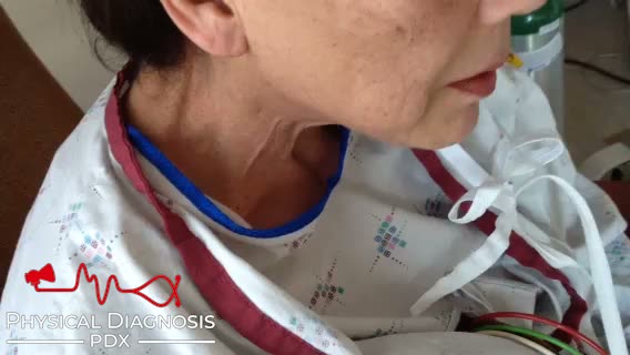
Kussmaul’s sign is defined as a paradoxical rise in jugular venous pressure during inspiration. Decreased intrathoracic pressure during inspiration normally leads to an increase in venous return to the right side of the heart, with an associated decrease in jugular venous pressure. When there is impaired filling of the right ventricle, the jugular veins instead become engorged. Causes of Kussmaul’s sign include right ventricular infarction, severe right ventricular failure, restrictive cardiomyopathy, constrictive pericarditis, and tricuspid stenosis.

Card Tutorial

Patient 2

This patient developed constrictive pericarditis many years after orthotopic heart transplantation, a rare sequala of that procedure. The astute observer will also note that in addition to Kussmaul’s sign, these videos demonstrate the presence of the W sign in the jugular venous waveform.

Video courtesy of BMJ case reports: “Jugular venous pulse in constrictive pericarditis”

Kussmaul’s Sign

Annotated

This patient also has

Right Ventricular Heave S4 Gallop
JVP Forearm Assessment Lancisi’s Sign

Teaching physical diagnosis through a library of real clinical findings.

  • About
  • Resources
  • Privacy
  • Acknowledgments