Physical Diagnosis PDX
Library
Cardiology Atlas Tutorial
Neurology Atlas Tutorial
Rheumatology Atlas Tutorial
Endocrinology Atlas Tutorial
Gastroenterology Atlas Tutorial
Pulmonology Atlas Tutorial
Infectious Disease Atlas Tutorial
Hematology-Oncology Atlas Tutorial
Nephrology Atlas Tutorial
Nails Atlas Tutorial
Hereditary Atlas Tutorial
Miscellaneous Atlas Tutorial
Cases Lectures Quizzes About
Library

Cardiology

Atlas Tutorial

Neurology

Atlas Tutorial

Rheumatology

Atlas Tutorial

Endocrinology

Atlas Tutorial

Gastroenterology

Atlas Tutorial

Pulmonology

Atlas Tutorial

Infectious Disease

Atlas Tutorial

Hematology-Oncology

Atlas Tutorial

Nephrology

Atlas Tutorial

Nails

Atlas Tutorial

Hereditary

Atlas Tutorial

Miscellaneous

Atlas Tutorial
Cases Lectures Quizzes About
  1. Home
  2. Cardiology
  3. Normal Jugular Venous Waveform

vimeo-472299681--Media-Quantitative-Assessment_Normal-Jugular-Venous-Pressure_JVP-Q-Tip-Video_cli.mp4

File is not under priv/media.

Patient 1 Patient 2 Patient 3 Patient 4 Patient 5
Clip 1 Clip 2 Clip 3 Clip 4
Compare
Previous patient Previous clip Next clip
Cardiology Jugular Venous Pulse

Normal Jugular Venous Waveform

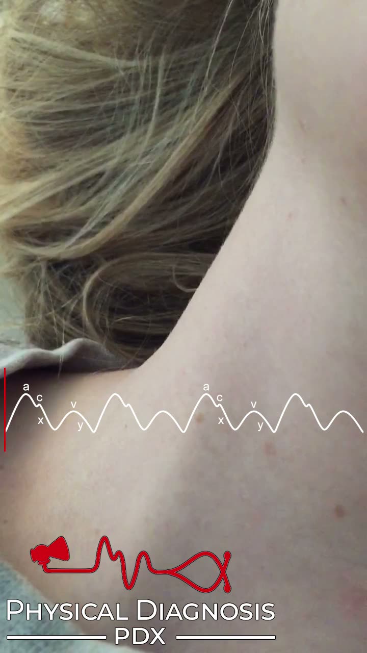
The normal jugular venous pulse is best evaluated via the right internal jugular vein, and is characterized by three peaks and two troughs. The first peak, called the a wave, results from atrial contraction during late diastole. Next, during early systole, ventricular contraction triggers closure of the tricuspid valve, producing the c wave (not visible at the bedside). In mid-systole, a combination of atrial relaxation and descent of the atrial floor during ventricular contraction results in the x descent. The third peak, the v wave, occurs as a result of atrial filling during late systole. Finally, passive ventricular filling in early diastole produces the y descent. The most conspicuous features of the venous pulse are the troughs, which generate obvious, aggressive inward movements of the skin. In the normal jugular venous waveform, the x descent is deeper than the y descent. Compared to the arterial pulse, the venous pulse involves a more diffuse area of the neck. It varies with patient positioning, respiratory cycle, and abdominal pressure (abdominojugular reflux). The normal venous pulse is not palpable. 

Card Tutorial

Patient 5

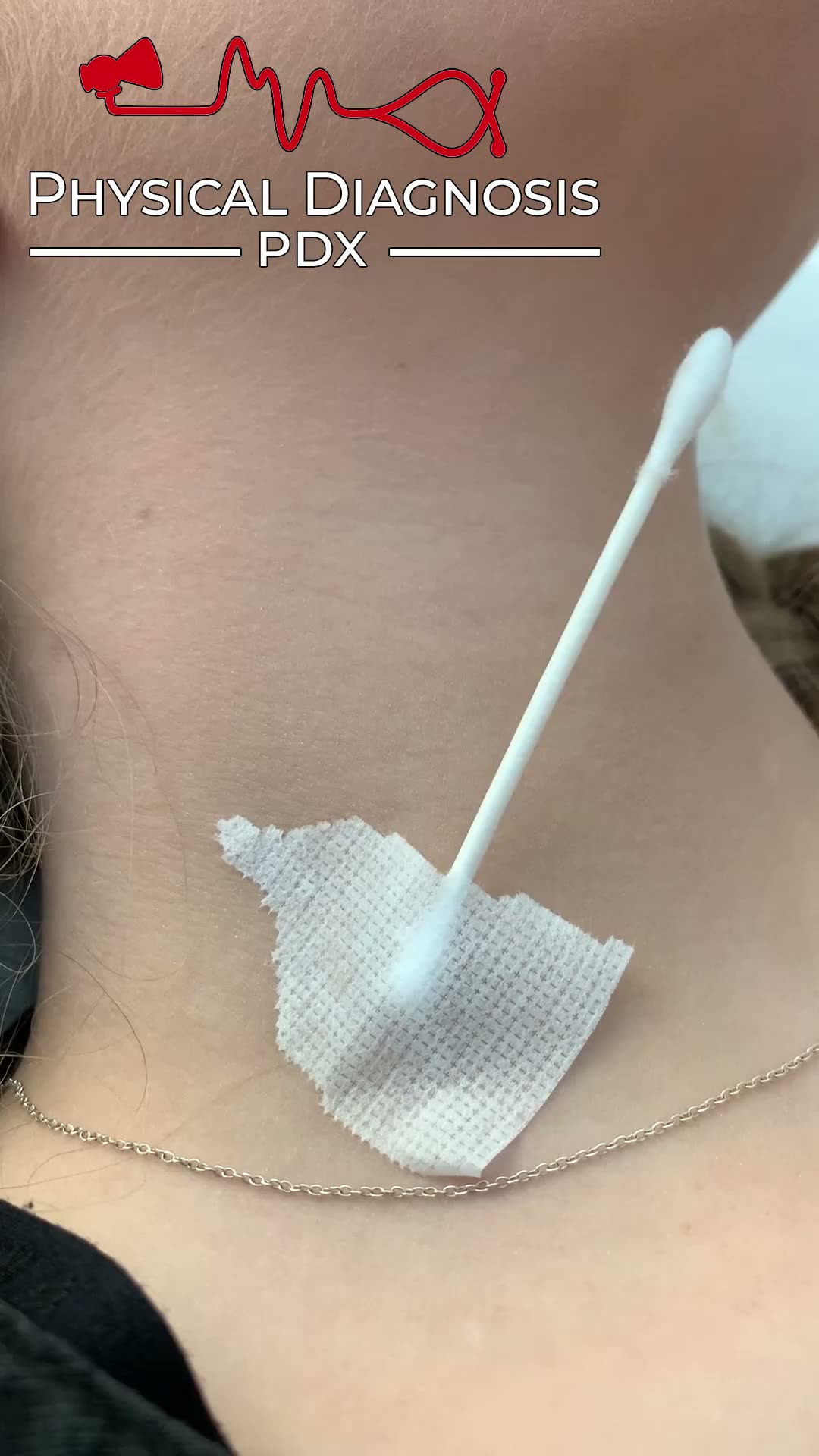
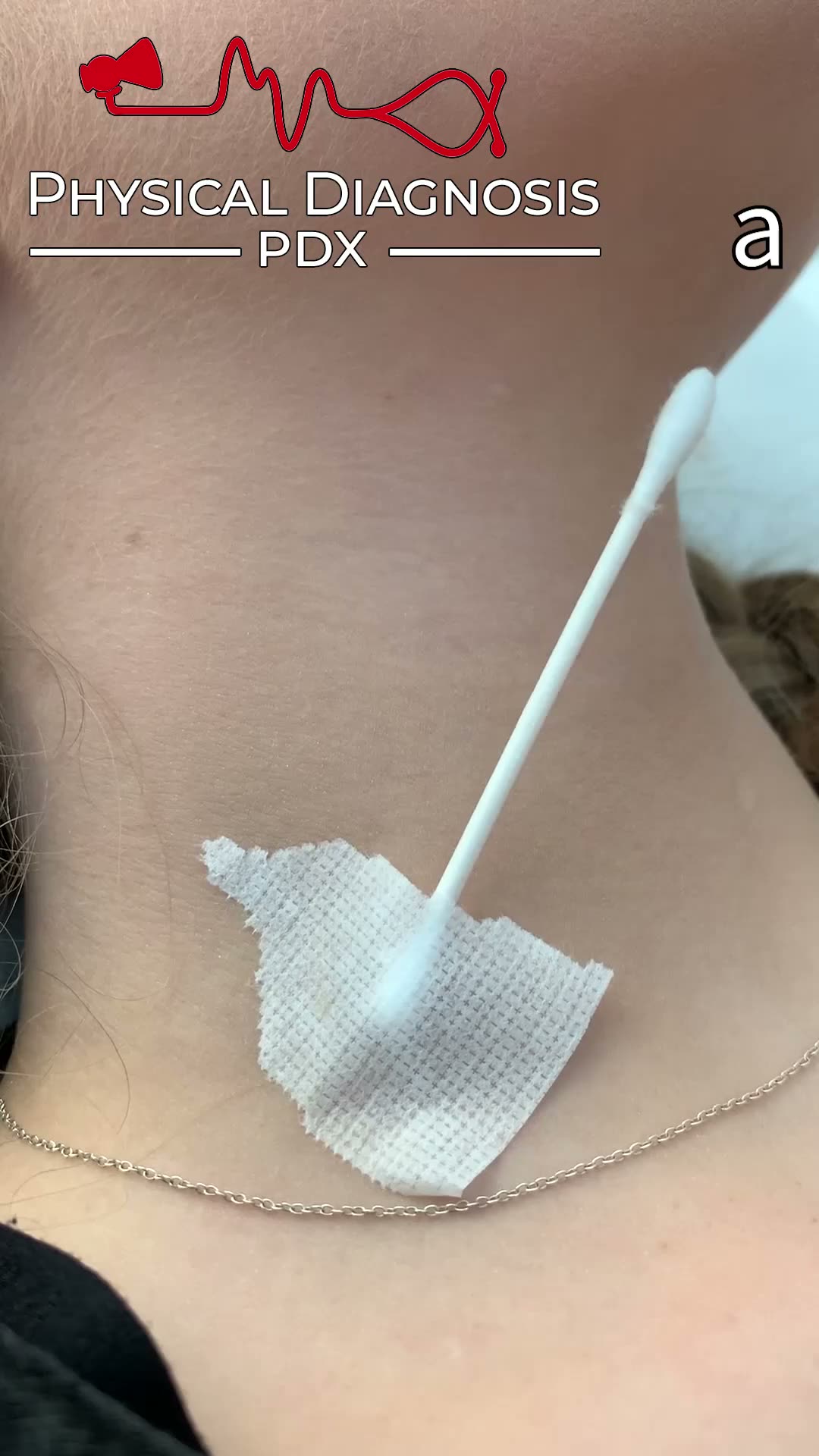
Here is a PDX-PDX team member demonstrating a normal jugular venous waveform. 

https://physicaldiagnosispdx.com/wp-content/uploads/JVP_Andre_-1.mp4

Normal Jugular Venous Pressure W Sign

Teaching physical diagnosis through a library of real clinical findings.

  • About
  • Resources
  • Privacy
  • Acknowledgments