Physical Diagnosis PDX
Library
Cardiology Atlas Tutorial
Neurology Atlas Tutorial
Rheumatology Atlas Tutorial
Endocrinology Atlas Tutorial
Gastroenterology Atlas Tutorial
Pulmonology Atlas Tutorial
Infectious Disease Atlas Tutorial
Hematology-Oncology Atlas Tutorial
Nephrology Atlas Tutorial
Nails Atlas Tutorial
Hereditary Atlas Tutorial
Miscellaneous Atlas Tutorial
Cases Lectures Quizzes About
Library

Cardiology

Atlas Tutorial

Neurology

Atlas Tutorial

Rheumatology

Atlas Tutorial

Endocrinology

Atlas Tutorial

Gastroenterology

Atlas Tutorial

Pulmonology

Atlas Tutorial

Infectious Disease

Atlas Tutorial

Hematology-Oncology

Atlas Tutorial

Nephrology

Atlas Tutorial

Nails

Atlas Tutorial

Hereditary

Atlas Tutorial

Miscellaneous

Atlas Tutorial
Cases Lectures Quizzes About
  1. Home
  2. Cardiology
  3. Split S1

vimeo-463373978--Annotated_SplitS1_2.mp4

File is not under priv/media.
Patient 1 Patient 2 Patient 3 Patient 4
Clip 1
Raw Compare
Next patient
Cardiology Heart Sounds

Split S1

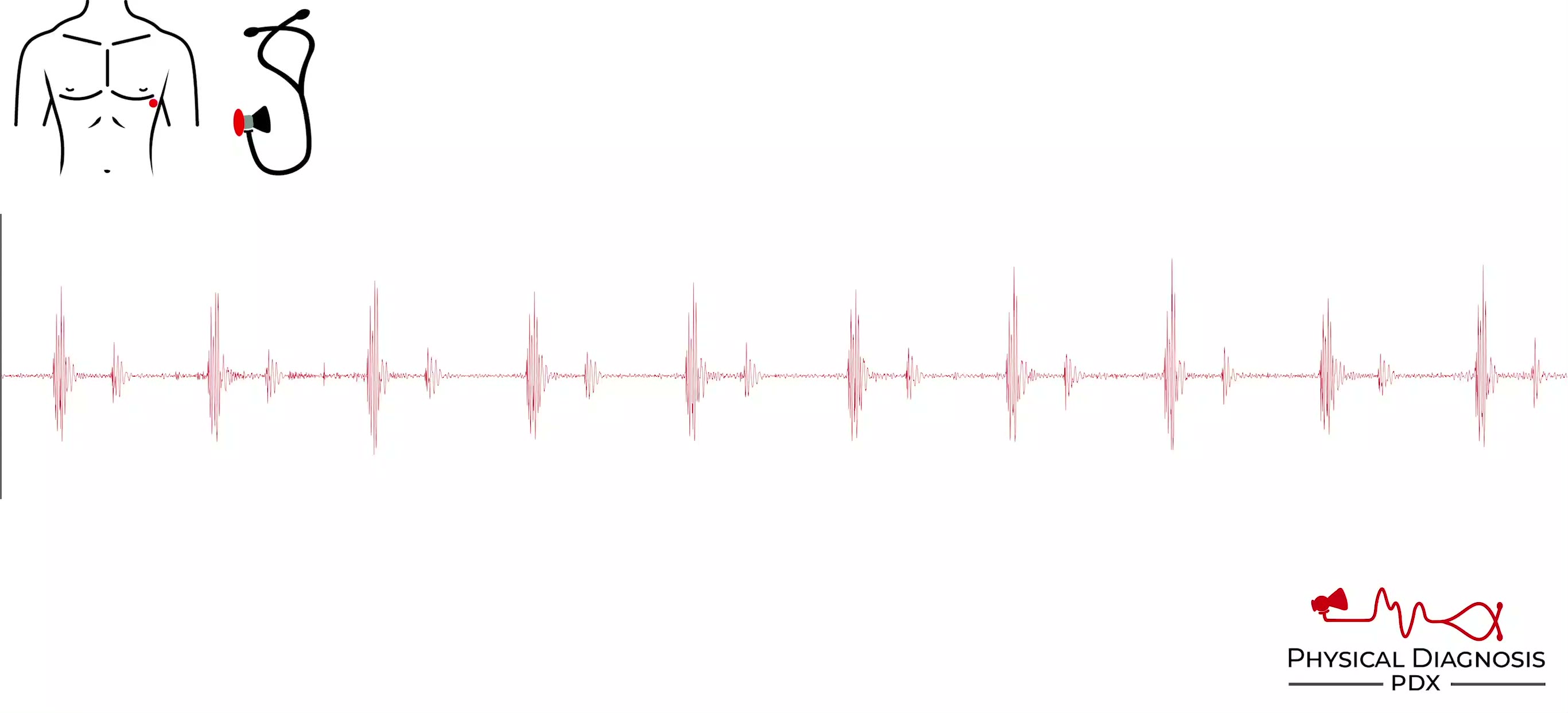
S1 is generated by closure of the mitral and tricuspid valves. These paired left- and right-sided valves do not close at exactly the same time, but the human ear cannot distinguish sounds that are 0.02-0.03 seconds apart as 2 separate sounds. When conditions further delay tricuspid valve closure, a split S1 can be appreciated. One of the most common causes of delayed tricuspid valve closure is a right bundle branch block.
Card Tutorial

Patient 1

This patient was admitted to the hospital for a non-cardiac reason and was noted to have a split S1. You can hear two sounds in quick succession followed by a single S2. There was no clear cause of the split S1 in this case.

Split S1

Annotated

Soft S1 Starr-Edwards Prosthetic Valve

Teaching physical diagnosis through a library of real clinical findings.

  • About
  • Resources
  • Privacy
  • Acknowledgments