Physical Diagnosis PDX
Library
Cardiology Atlas Tutorial
Neurology Atlas Tutorial
Rheumatology Atlas Tutorial
Endocrinology Atlas Tutorial
Gastroenterology Atlas Tutorial
Pulmonology Atlas Tutorial
Infectious Disease Atlas Tutorial
Hematology-Oncology Atlas Tutorial
Nephrology Atlas Tutorial
Nails Atlas Tutorial
Hereditary Atlas Tutorial
Miscellaneous Atlas Tutorial
Cases Lectures Quizzes About
Library

Cardiology

Atlas Tutorial

Neurology

Atlas Tutorial

Rheumatology

Atlas Tutorial

Endocrinology

Atlas Tutorial

Gastroenterology

Atlas Tutorial

Pulmonology

Atlas Tutorial

Infectious Disease

Atlas Tutorial

Hematology-Oncology

Atlas Tutorial

Nephrology

Atlas Tutorial

Nails

Atlas Tutorial

Hereditary

Atlas Tutorial

Miscellaneous

Atlas Tutorial
Cases Lectures Quizzes About
  1. Home
  2. Cardiology
  3. W Sign

vimeo-472318229--Media-Qualitative-Assessment_Kussmauls-Sign_JVP_Kussmauls-2.mp4

File is not under priv/media.
Patient 1
Clip 1 Clip 2
Compare
Next clip
Cardiology Jugular Venous Pulse

W Sign

The normal jugular venous waveform contains two descents, x and y. Under normal conditions, the x descent is more prominent than the y descent. In constrictive pericarditis, the unyielding pericardium causes elevated atrial pressure and limits ventricular filling to the early diastolic period, resulting in a sharp and deep y descent, known as Friedreich’s sign. In some patients, the prominent y descent occurs in combination with a prominent x descent, creating two steep troughs known as the “W” sign. This finding is helpful in distinguishing constrictive pericarditis from restrictive cardiomyopathy, in which the x descent is typically diminished.

Card Tutorial

Patient 1

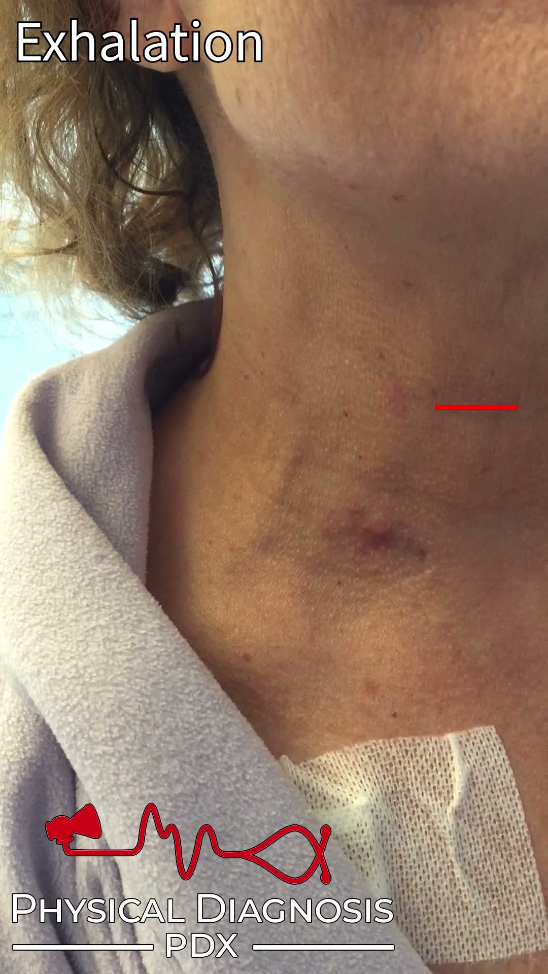
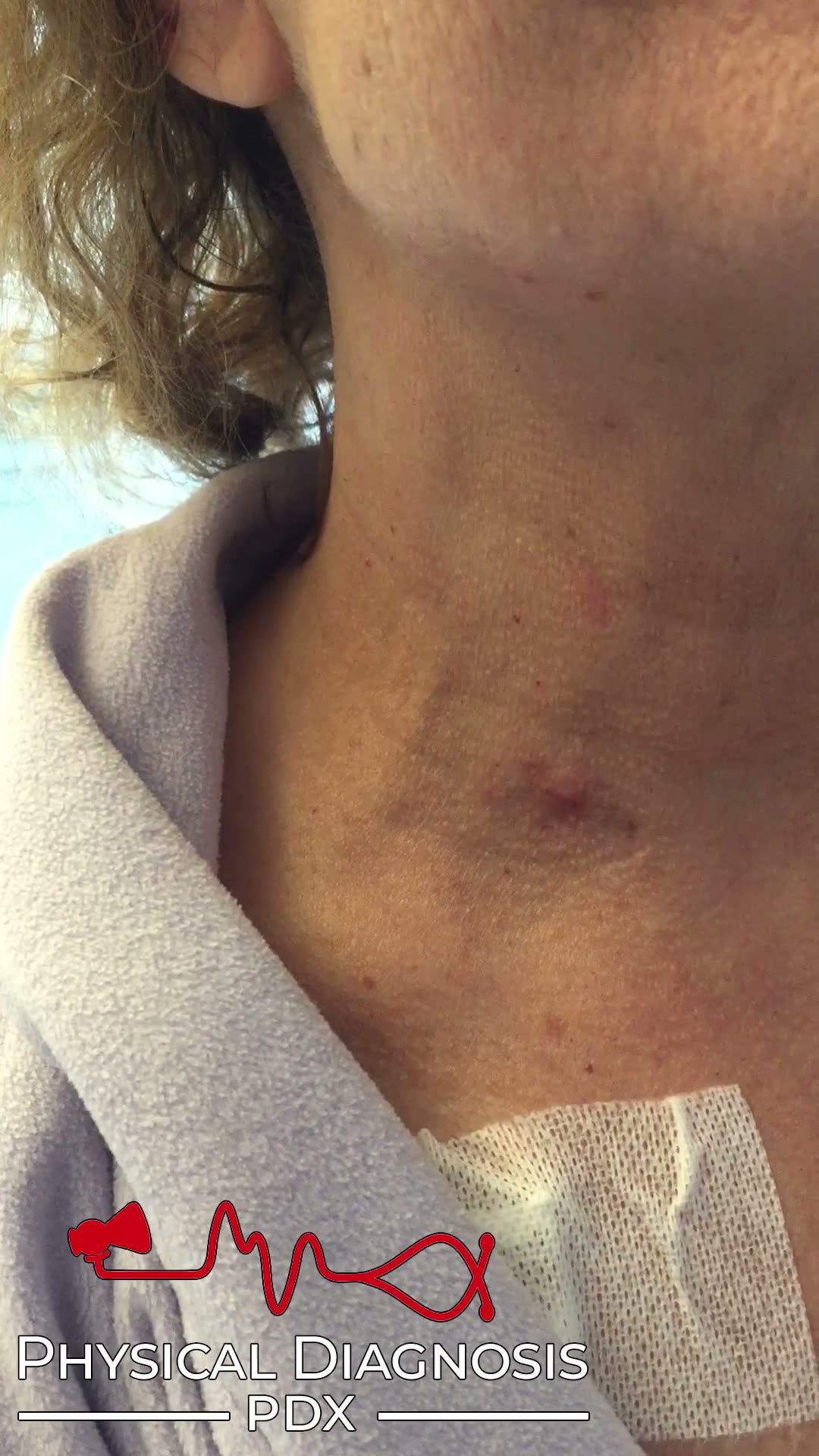
This patient developed constrictive pericarditis many years after orthotopic heart transplantation, a rare sequala of that procedure. These videos demonstrate sharp and deep x and y descents of the jugular venous waveform. The astute observer will also note the presence of Kussmaul’s sign.

Video courtesy of BMJ case reports: “Jugular venous pulse in constrictive pericarditis”

*It may be easier to appreciate this video by slowing it down to half speed (click the gear icon in the bottom right corner of the video)

W sign

Annotated

Normal Jugular Venous Waveform

Teaching physical diagnosis through a library of real clinical findings.

  • About
  • Resources
  • Privacy
  • Acknowledgments