Physical Diagnosis PDX
Library
Cardiology Atlas Tutorial
Neurology Atlas Tutorial
Rheumatology Atlas Tutorial
Endocrinology Atlas Tutorial
Gastroenterology Atlas Tutorial
Pulmonology Atlas Tutorial
Infectious Disease Atlas Tutorial
Hematology-Oncology Atlas Tutorial
Nephrology Atlas Tutorial
Nails Atlas Tutorial
Hereditary Atlas Tutorial
Miscellaneous Atlas Tutorial
Cases Lectures Quizzes About
Library

Cardiology

Atlas Tutorial

Neurology

Atlas Tutorial

Rheumatology

Atlas Tutorial

Endocrinology

Atlas Tutorial

Gastroenterology

Atlas Tutorial

Pulmonology

Atlas Tutorial

Infectious Disease

Atlas Tutorial

Hematology-Oncology

Atlas Tutorial

Nephrology

Atlas Tutorial

Nails

Atlas Tutorial

Hereditary

Atlas Tutorial

Miscellaneous

Atlas Tutorial
Cases Lectures Quizzes About
  1. Home
  2. Cardiology
  3. Giant a Wave

vimeo-472319427--Media-Qualitative-Assessment_Giant-A-Wave_JVP_Giant-A-Wave-2_Annotated.mp4

File is not under priv/media.
Patient 1 Patient 2 Patient 3 Patient 4 Patient 5 Patient 6 Patient 7
Clip 1
Previous patient Next patient
Cardiology Jugular Venous Pulse

Giant a Wave

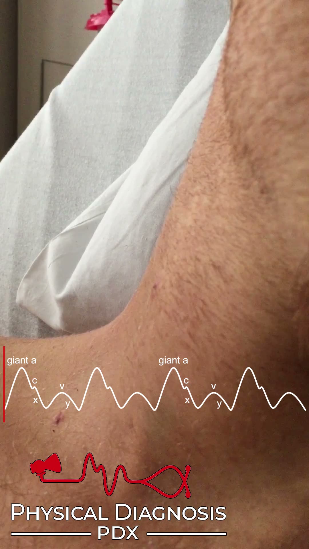
The a wave of the jugular venous waveform occurs as a result of right atrial contraction. When there is resistance to right ventricular filling, right atrial contraction generates increased pressure, resulting in giant a waves. These waves occur with every beat, unlike cannon a waves, which usually occur intermittently. Causes of giant a waves include tricuspid stenosis and increased right ventricular end-diastolic pressure from any cause (e.g., pulmonary hypertension).

Card Tutorial

Patient 2

This patient was admitted to the hospital with decompensated right heart failure from severe pulmonary hypertension caused by methamphetamine use. On examination, there was a a right ventricular heave as well as a loud pulmonary component of the second heart sound, consistent with pulmonary hypertension. These videos demonstrate a jugular venous waveform with giant a waves. Pulmonary hypertension creates resistance to right atrial filling as a result of elevated right ventricular end-diastolic pressure.

*It may be easier to appreciate this video by slowing it down to half speed (click the gear icon in the bottom right corner of the video)

Giant a Wave

Annotated

Friedreich’s Sign Hand Vein Assessment

Teaching physical diagnosis through a library of real clinical findings.

  • About
  • Resources
  • Privacy
  • Acknowledgments