Cardiology
JVP, pulses, auscultation, and the cardiac exam. 83 findings
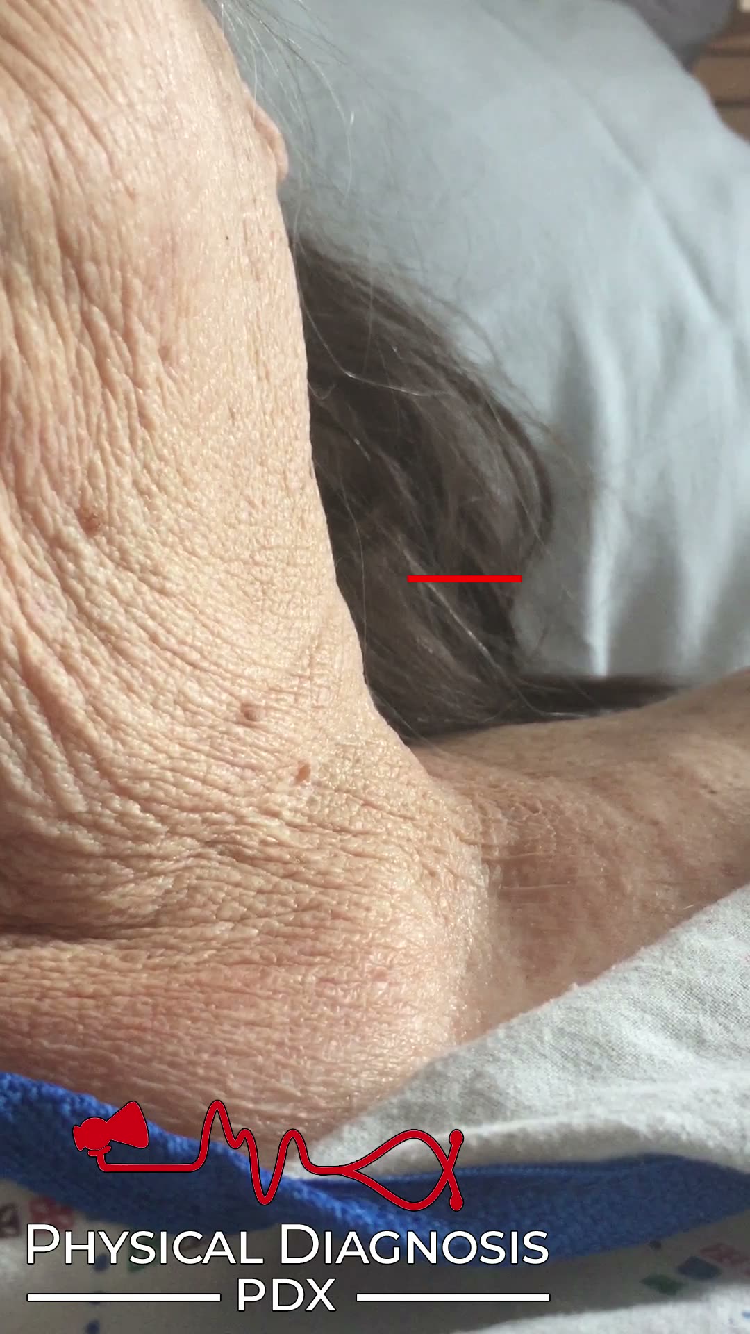
Abdominojugular Reflux
2 patients · 4 clips
The abdominojugular (reflux) test is a physical examination maneuver that is performed by placing firm pressure over the patient’s abdomen with the palm of the hand while observing the effect on jugular venous pressure.
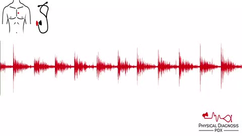
Aortic Regurgitation
13 patients · 38 clips
- Soft, decrescendo murmur that starts right at S2 and trails off (turbulence is highest in early diastole when the pressure gradient between the aorta and LV is highest). - Murmur may be best heard with the patient lean
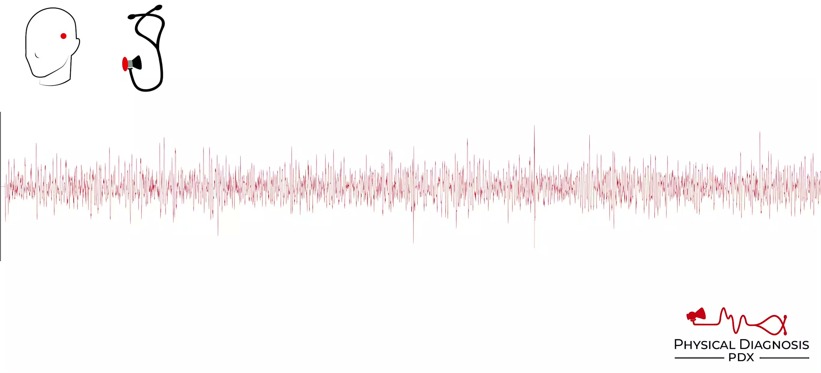
Carotid-cavernous Fistula
1 patients · 1 clips
Carotid-cavernous fistula describes the abnormal communication between the carotid artery and the cavernous sinus. It is most often a complication from head trauma resulting in a basilar skull fracture. A continuous brui
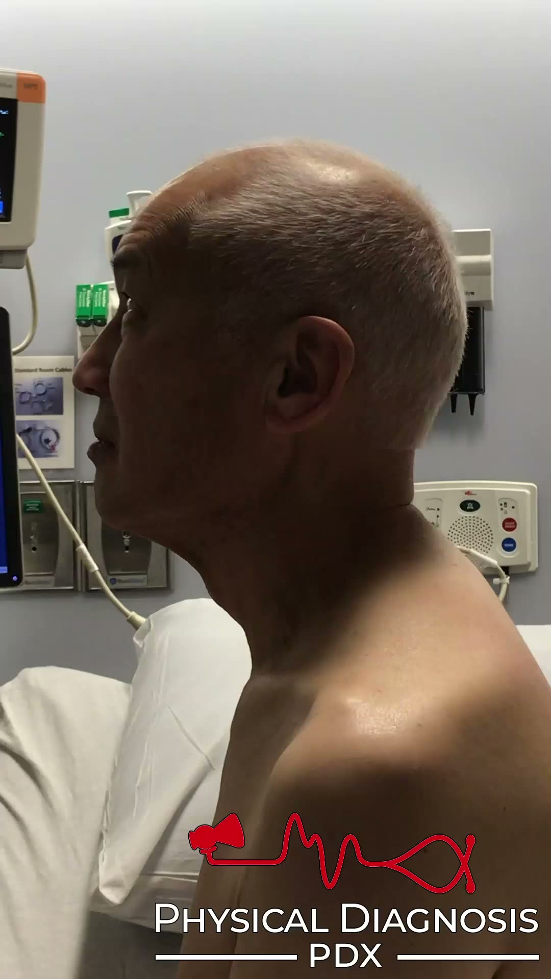
De Musset’s Sign
1 patients · 1 clips
Forward and back head bob seen in aortic regurgitation Card Tutorial Patient 1: This is an older man with an aortic root aneurysm complicated by moderate to severe aortic regurgitation. This patient also has a decrescend
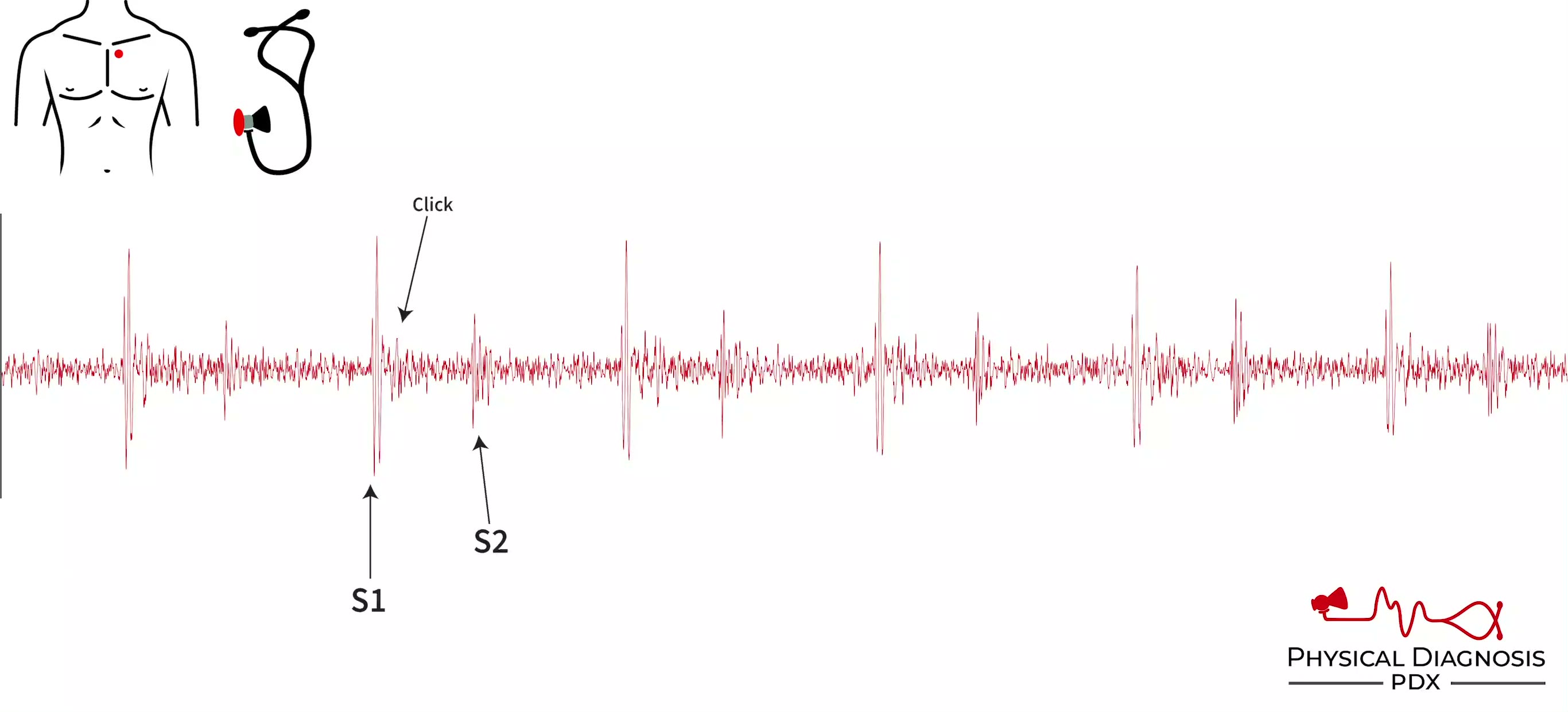
Ejection Click
5 patients · 10 clips
An ejection click is an early systolic sound, and occurs when an abnormal aortic or pulmonic valve (eg, bicuspid aortic valve) opens, or from ejection of blood into a dilated great vessel (aorta or pulmonary artery, such
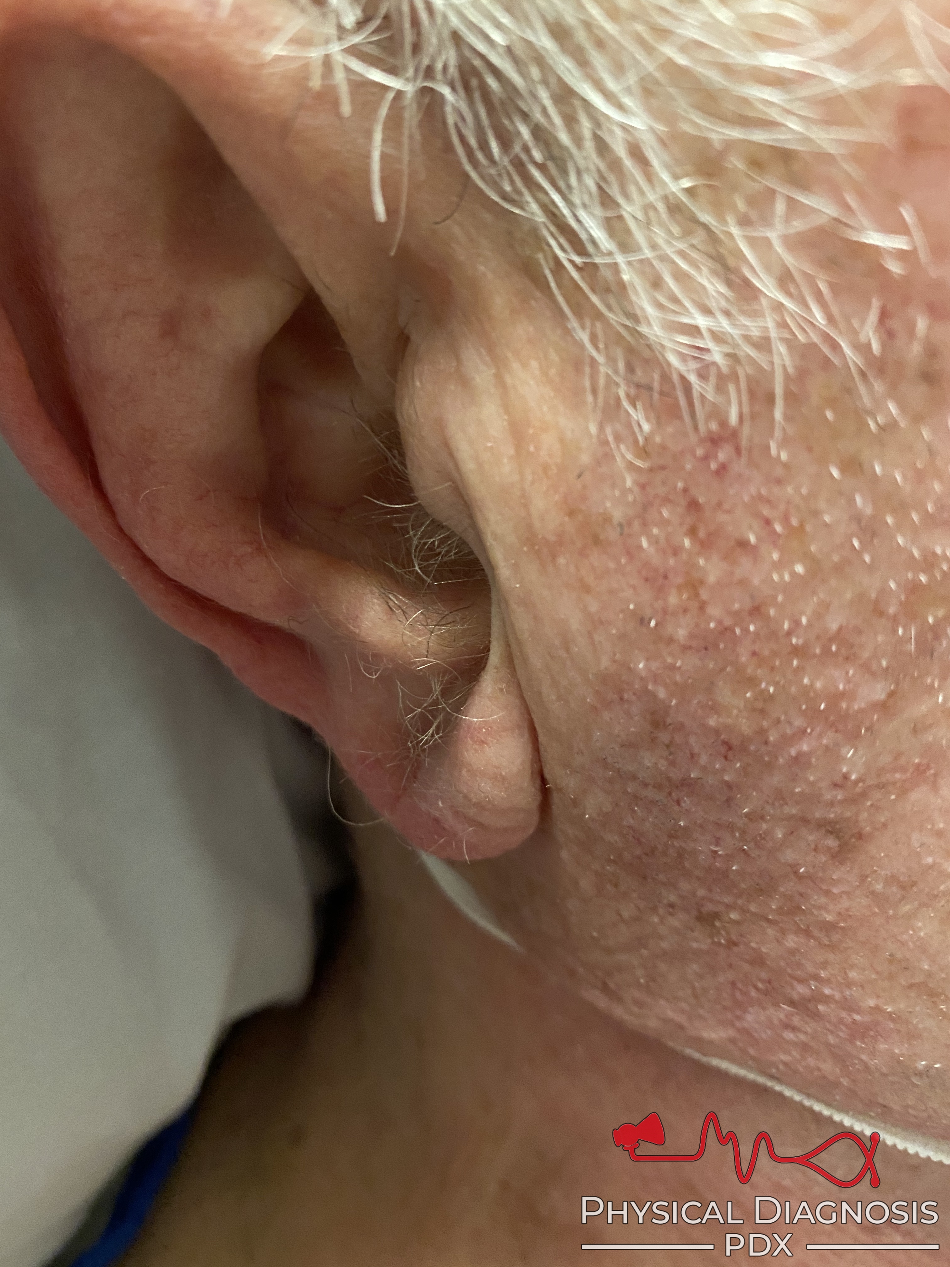
Frank’s Sign
2 patients · 3 clips
Diagonal earlobe crease, associated with coronary artery disease. Card Tutorial Patient 1: This patient has Frank’s sign. Patient 2: This patient has Frank’s sign.
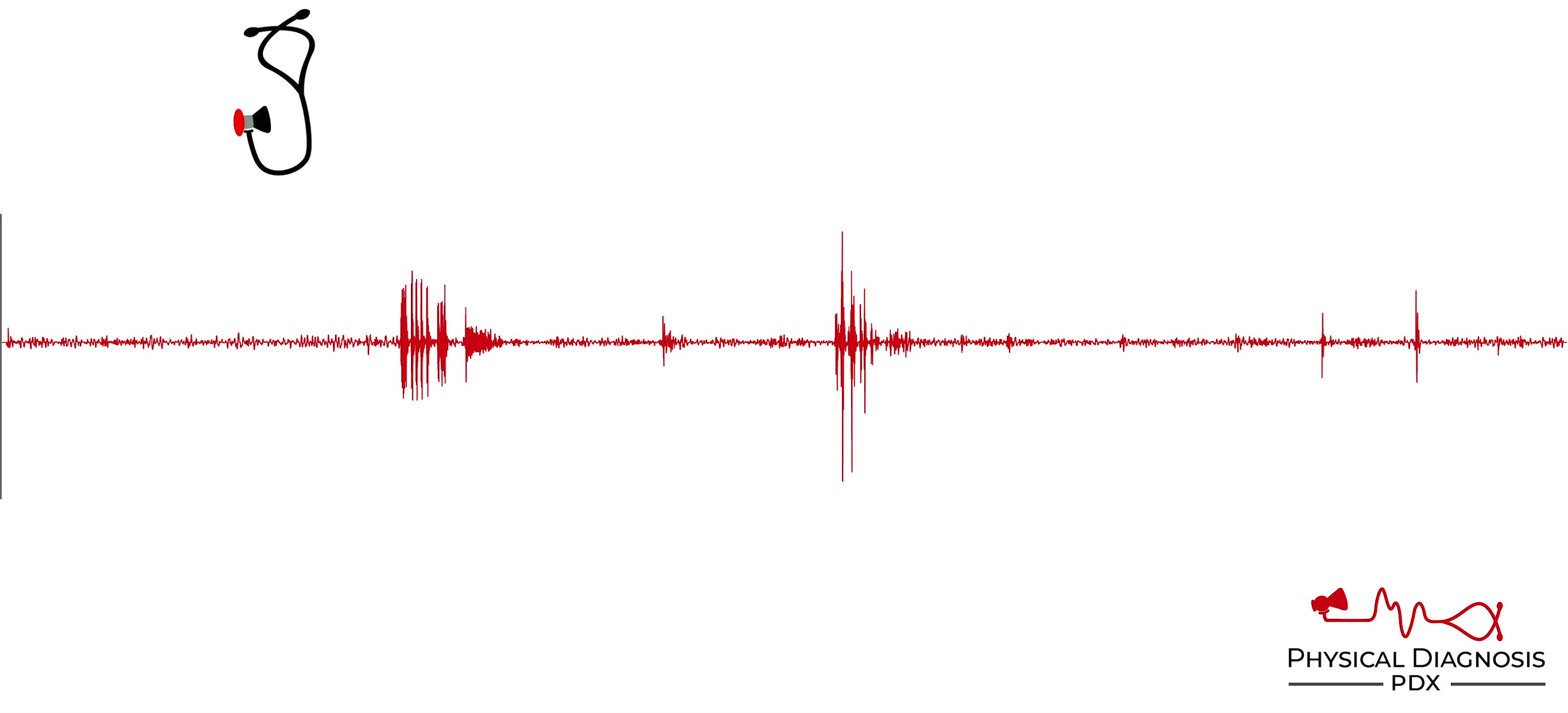
High Pitched Bowel
1 patients · 2 clips
Patient 1 This is an older woman with recurrent small bowel obstruction of unknown etiology, admitted for another bout. Note the high-pitched “tinkle” sounds.
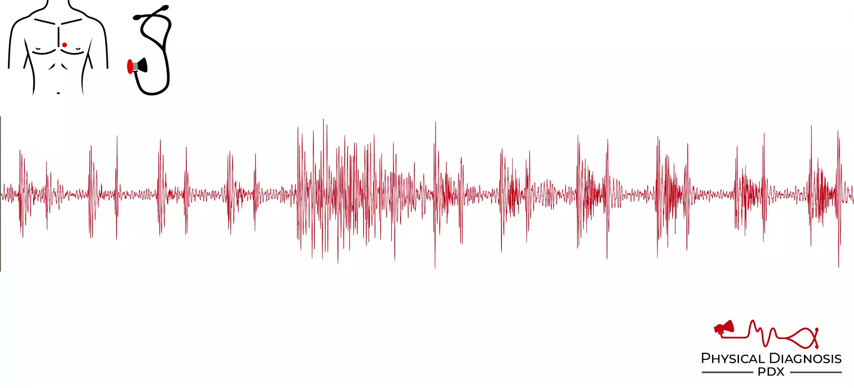
Hypertrophic Obstructive Cardiomyopathy (HOCM)
2 patients · 4 clips
The HOCM murmur is louder when the ventricular volume is low, as the outflow tract is narrower, so you can make this murmur louder by having the patient Valsalva or go from squatting to standing. The murmur will become s

Lateral Head Bob
4 patients · 7 clips
Lateral head bob seen in tricuspid regurgitation. Card Tutorial Patient 1 This patient has severe tricuspid regurgitation. Notice the side-to-side head bob. This patient also has Lancisi’s sign. Patient 2 This patient ha
Mechanical S2
8 patients · 23 clips
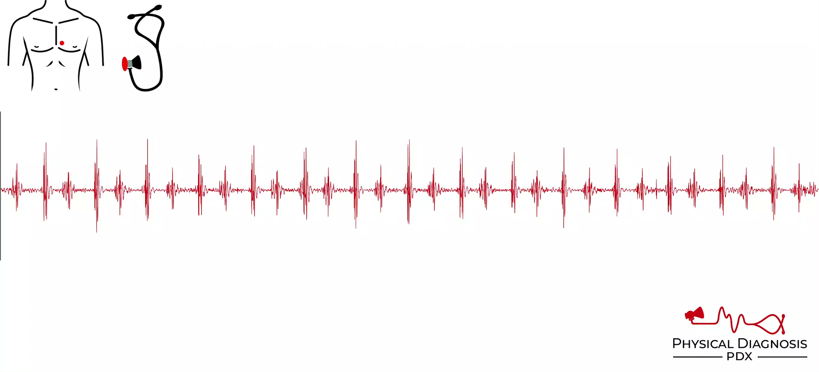
A mechanical S2 is generated by closure of a prosthetic heart valve (usually in the aortic position). It has a “metallic” quality. Mechanical aortic valves can often be heard with the naked ear. Card Tutorial Patient 1:
Mitral Stenosis
1 patients · 4 clips
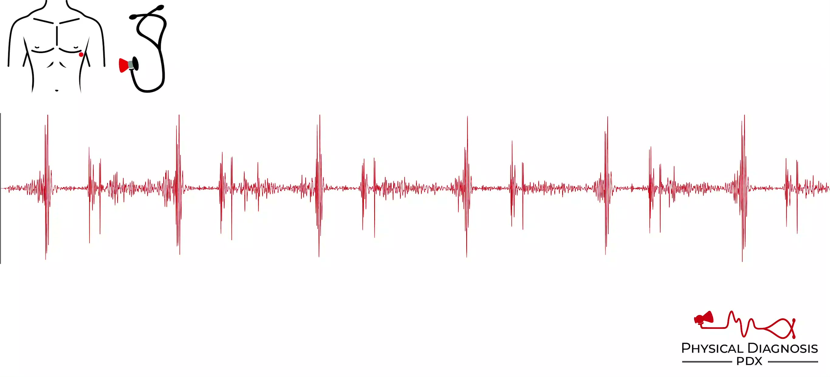
Mitral stenosis causes a low pitched, rumbling, diastolic murmur after the opening snap that continues through diastole and gets louder (presystolic accentuation) just before S1 due to atrial contraction best heard with
Mitral Valve Prolapse
4 patients · 8 clips
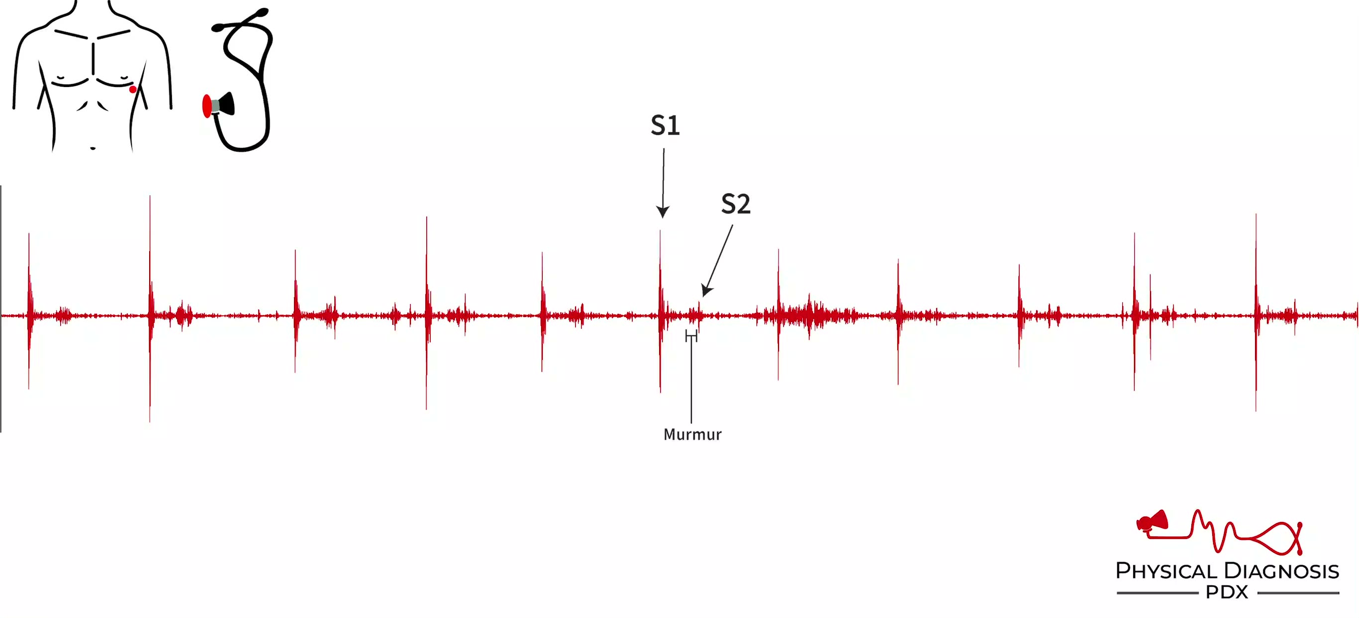
- Mitral valve prolapse is a variant of MR in which some part of the mitral apparatus is too long, so that the valve does not function well at low volumes. At higher volumes, the valve leaflet may coapt normally. At low
Persistently Split S2
6 patients · 10 clips
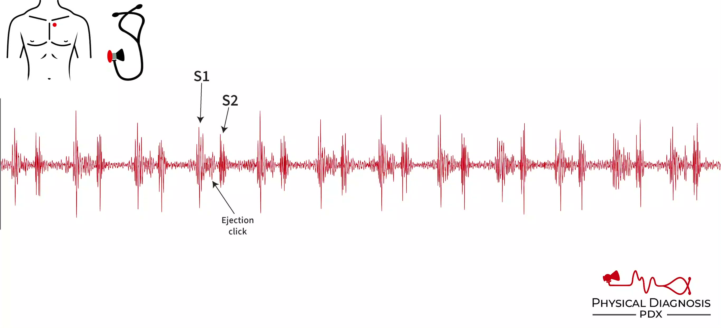
A persistently split S2 describes the presence of a split S2 on expiration that widens further upon inspiration. The most common cause of a persistently split S2 is a right bundle branch block, but it can occur whenever
Right Ventricular Heave
1 patients · 1 clips
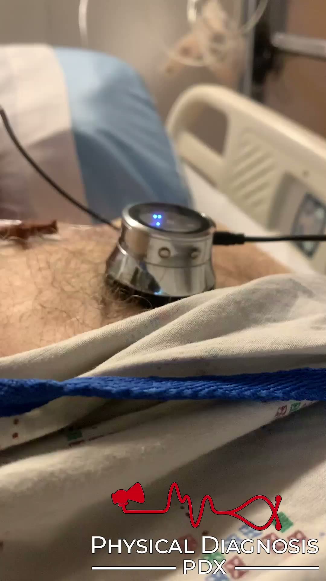
Visible or palpable chest wall impulses, known as heaves/lifts, may be from a variety of causes such as valvular regurgitation, pressure overload, or cardiac aneurysms. Card Tutorial Patient 1: This is an older man who w
Vegetation Plop
1 patients · 2 clips
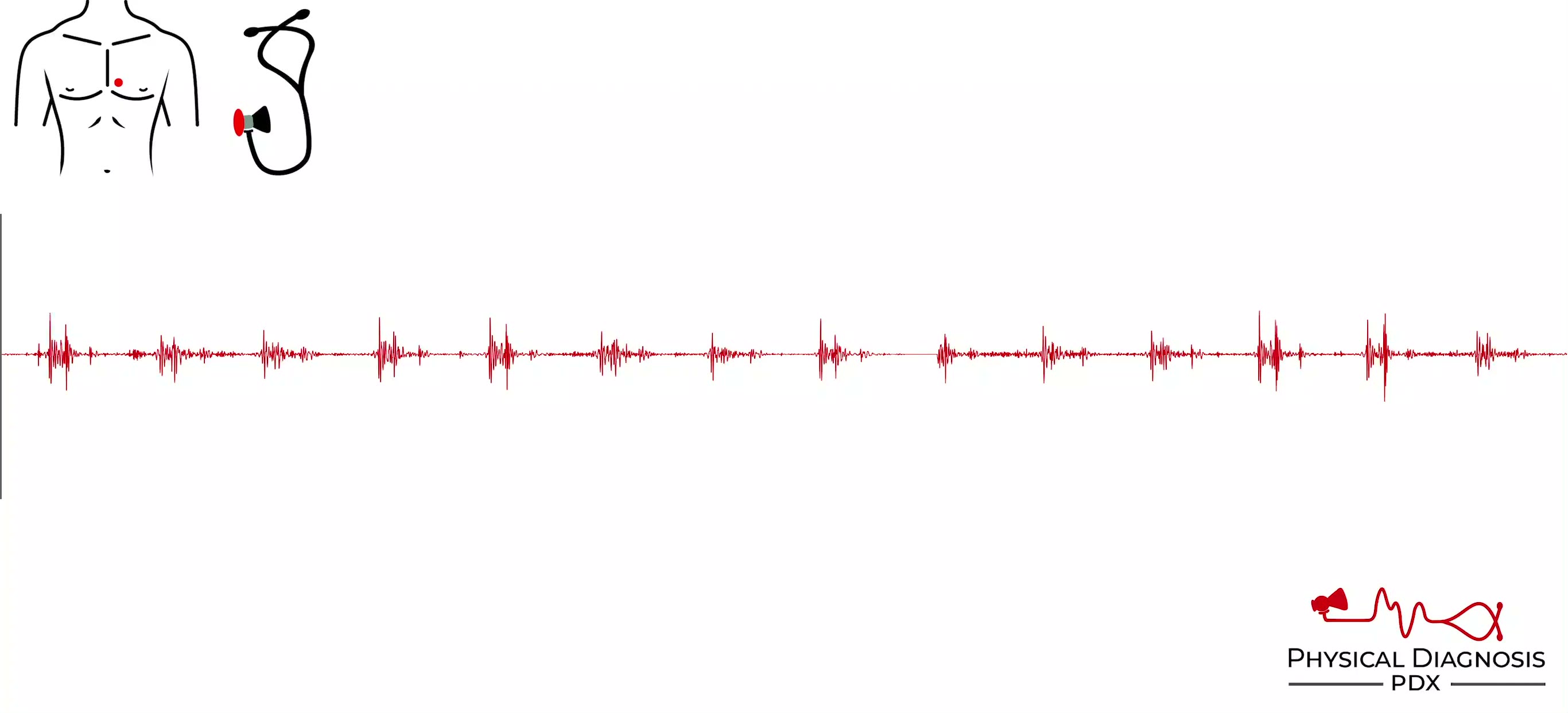
The vegetation plop is similar to the tumor plop, but occurs as a result of large vegetation from endocarditis. Card Tutorial Patient 1: This was a middle-aged man who was admitted to the hospital with infective endocard
Aortic Stenosis
9 patients · 25 clips
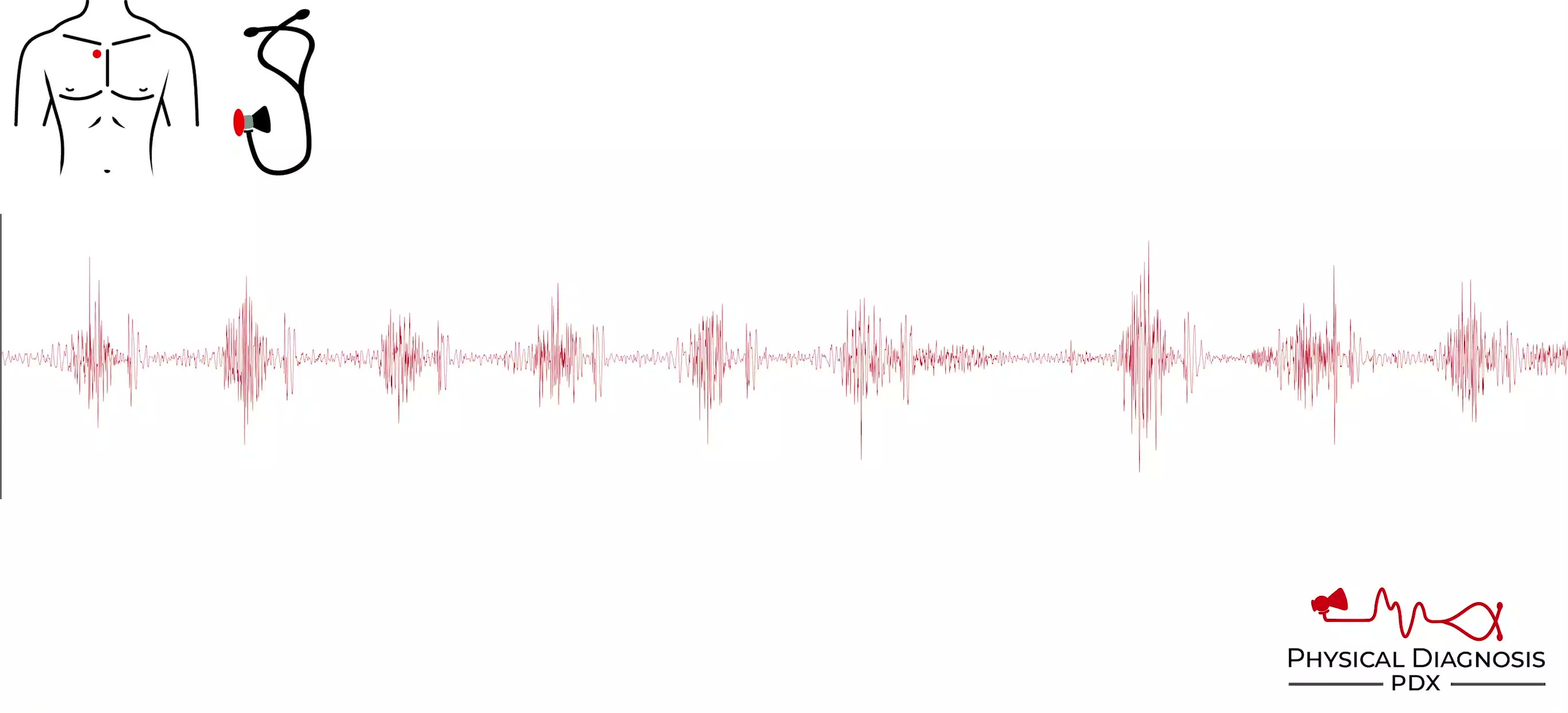
This murmur is systolic, crescendo-decrescendo in shape, harsh in quality, located in the sash distribution (clavicle to apex), and radiates to clavicle or neck. Causes are usually calcification in older patients (age >
Auscultory Alternans
1 patients · 1 clips
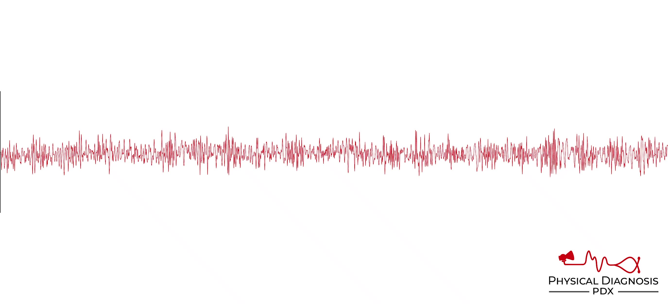
Clinical finding with teaching media.
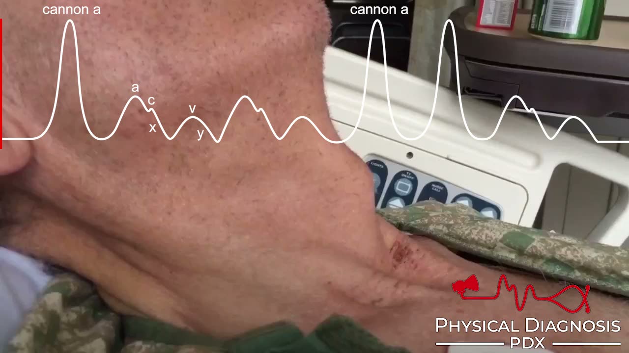
Cannon a Wave
2 patients · 4 clips
The a wave of the jugular venous waveform occurs as a result of right atrial contraction. When there is resistance to right ventricular filling, right atrial contraction generates increased pressure, resulting in large a
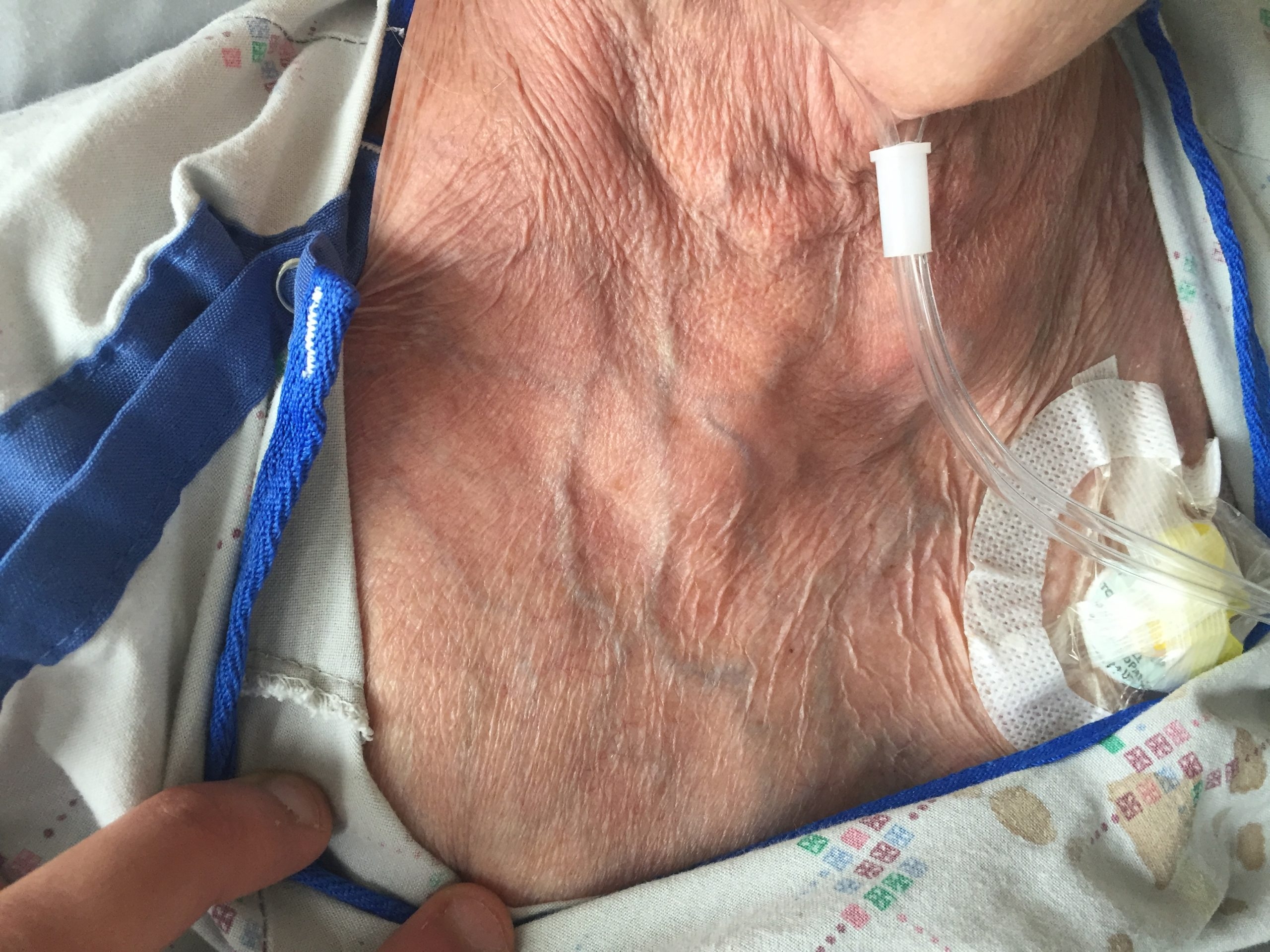
Dilated Veins On Chest
1 patients · 2 clips
Superior vena cava (SVC) syndrome occurs when obstruction of the SVC results in impaired cardiac filling, giving rise to a constellation of characteristic symptoms and signs. Dyspnea is one of the most frequent symptoms;
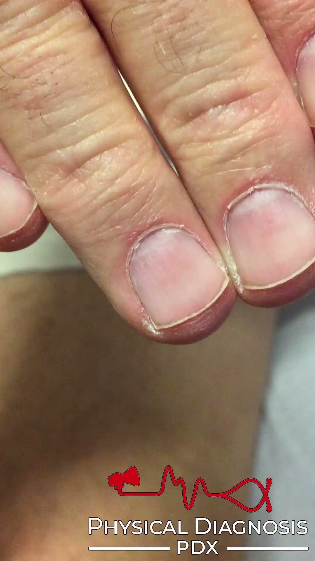
Quincke’s Pulse
10 patients · 10 clips
Pulsation of nail beds. Card Tutorial Patient 1: This is an older man with moderate alcohol use who developed the clinical syndrome of heart failure. Echocardiography was unremarkable. He was noted to have a wide pulse p
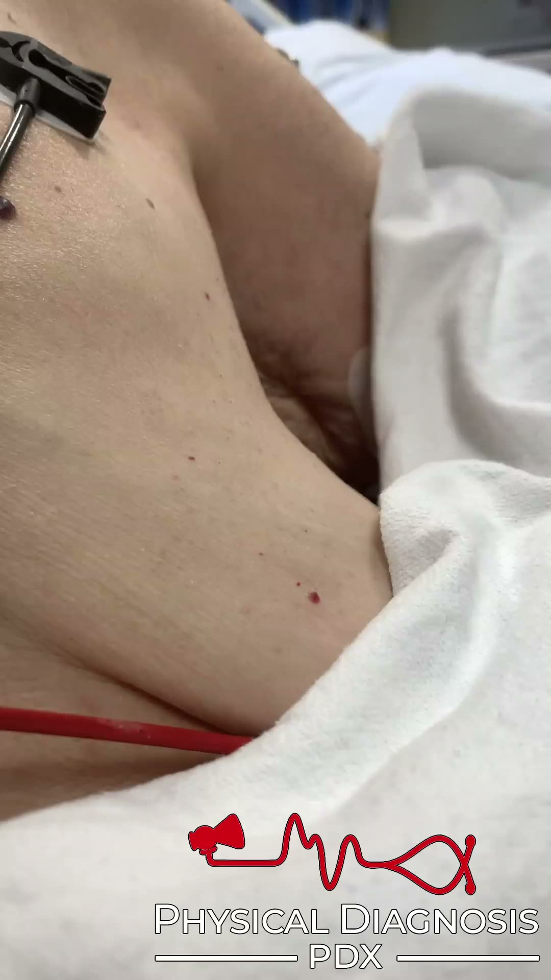
Regurgitant Lesion
1 patients · 1 clips
Visible or palpable chest wall impulses, known as heaves/lifts, may be from a variety of causes such as valvular regurgitation, pressure overload, or cardiac aneurysms. Card Tutorial Patient 1: This is an older woman wit
Aorto-Right Ventricular Fistula
1 patients · 2 clips
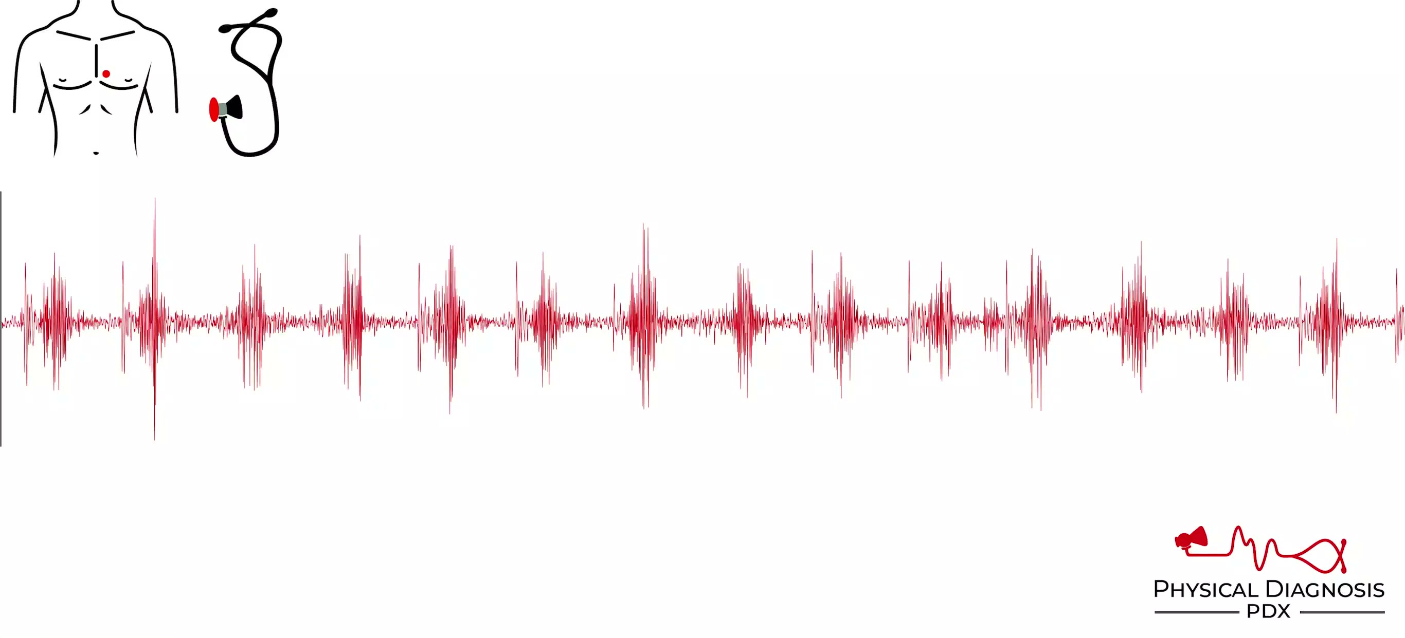
Card Tutorial Patient 1: This patient has
Cardiac Tamponade
1 patients · 2 clips
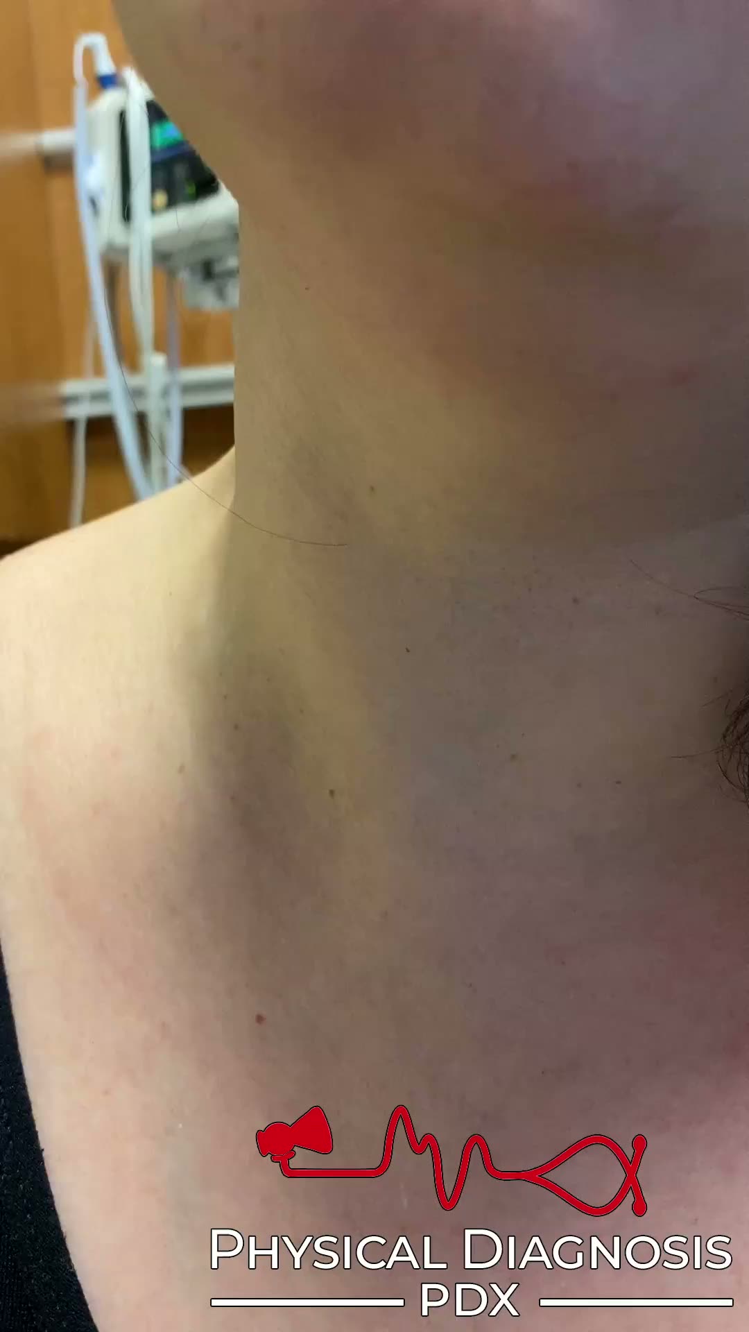
In the setting of cardiac tamponade, the jugular venous pulse is elevated. Qualitatively, the y descent is diminutive or absent. Card Tutorial Patient 1: This is a previously healthy young woman who was admitted to the h
Electronic Hum Left Ventricular Assist Device
1 patients · 1 clips
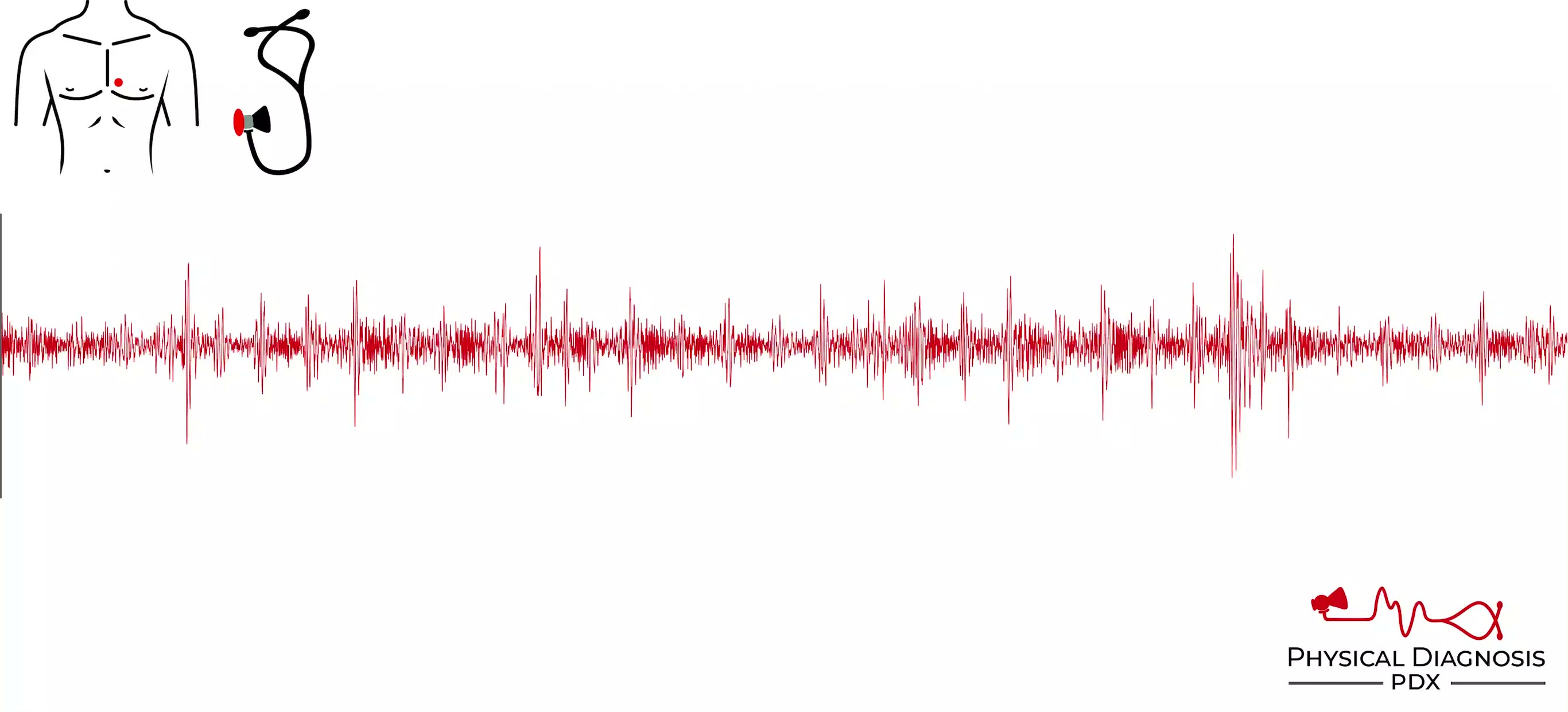
Patient 1: This is a middle-aged man with nonischemic cardiomyopathy who underwent left ventricular assist device placement. Notice the electronic hum of the device. This patient also has diffuse inspiratory rales though
Enlarged Forehead Veins
1 patients · 2 clips
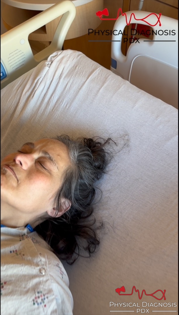
This is a middle-aged woman with methamphetamine-induced cardiomyopathy. Note how the forehead veins engorge when she is reclined. This is caused by venous congestion and can be an immediate clue the presence of elevated
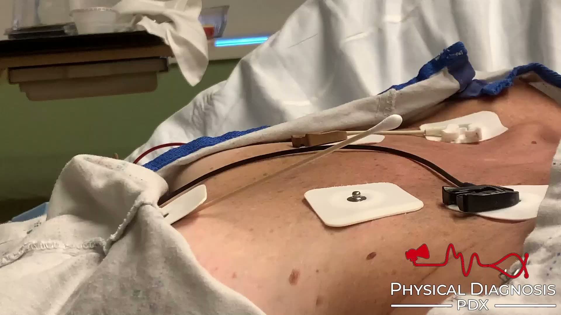
Gallop
2 patients · 4 clips
Pulmonology Tutorial Patient 1: This is a young woman with non-ischemic cardiomyopathy related to methamphetamine use, who was admitted to the hospital for decompensated heart failure. She was found to have a right-sided
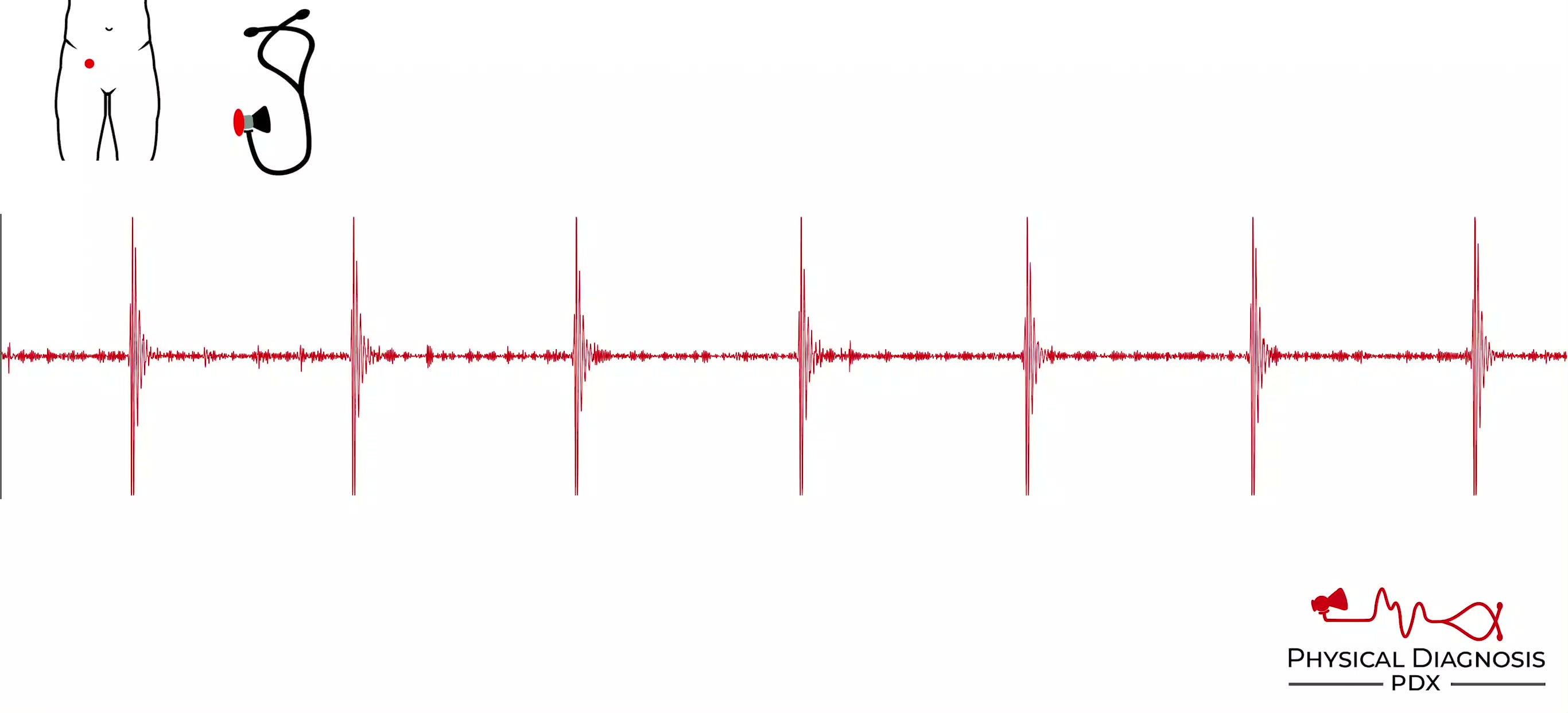
Traube’s Sign
1 patients · 2 clips
Pistol shot sounds heard on auscultation over arterial pulse in aortic regurgitation. Card Tutorial Patient 1: This is a middle-aged man with active intravenous drug use who was found to have infective endocarditis of th
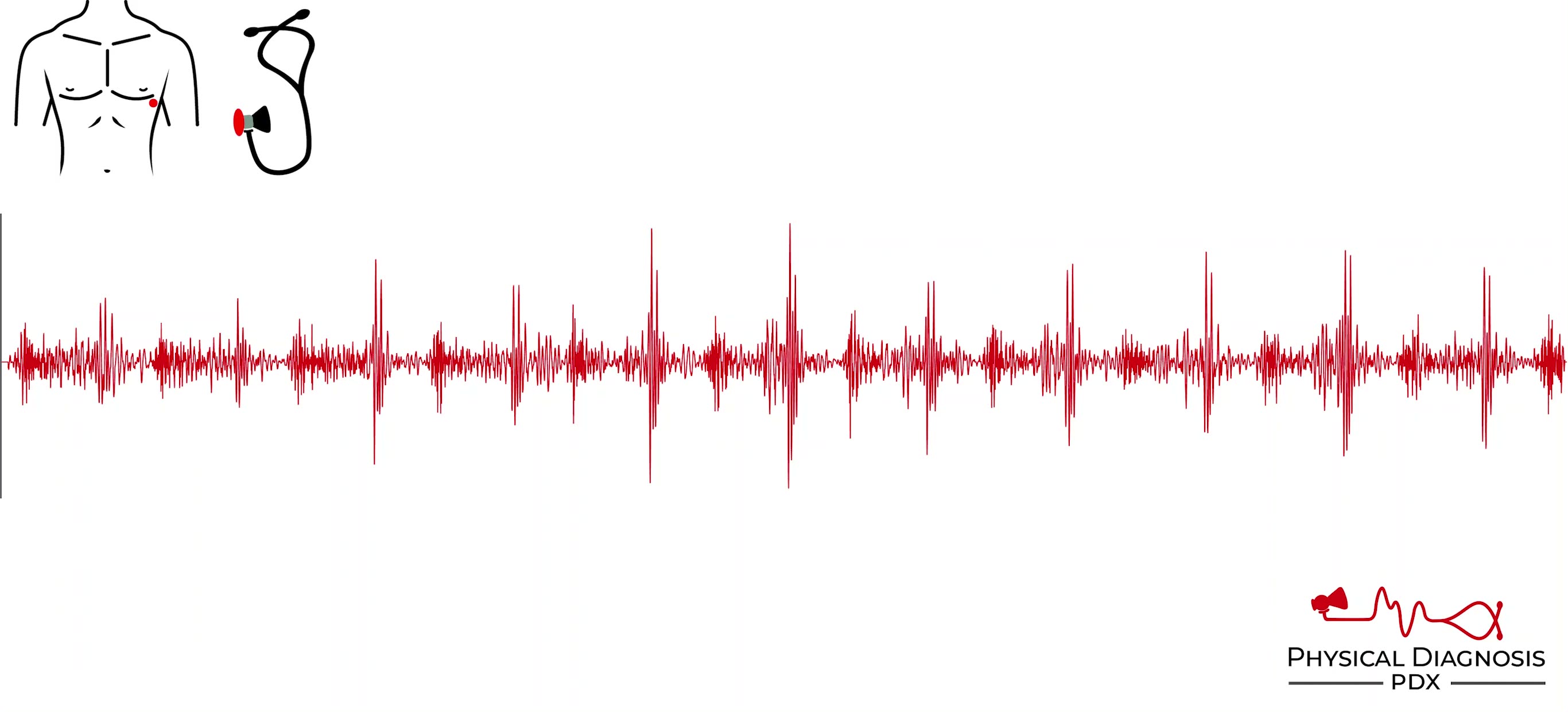
Austin Flint Murmur
2 patients · 8 clips
Aortic Regurgitation any be associated with a low-pitched, “blubbering”, mid-to-late diastolic murmur heard over the apex, known as the Austin Flint murmur. The mechanism is thought to be distortion and early closure of
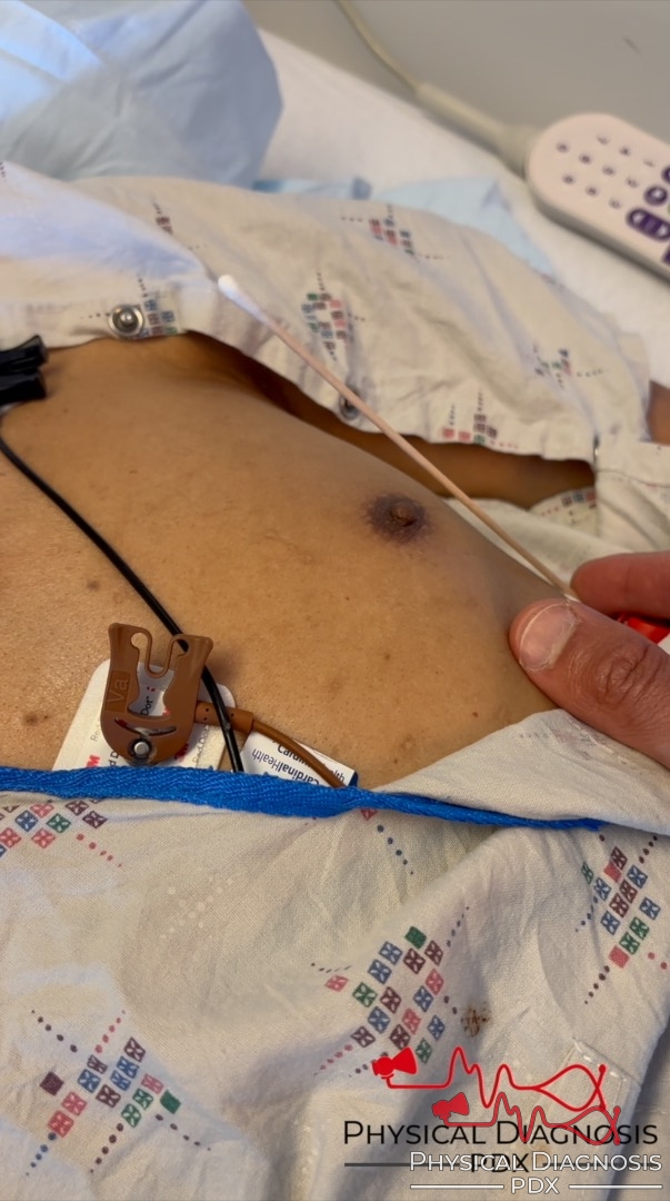
Displaced Point of Maximal Impulse
1 patients · 2 clips
**Patient 1:** This is an older man with familial cardiomyopathy, admitted with cardiogenic shock. Note the laterally displaced point of maximal impulse. You can also appreciate an extra bounce in the Q-tip from a gallop
Elevated Jugular Venous Pressure
11 patients · 19 clips
The traditional “window” for viewing the jugular venous pulse is confined to the region of the neck between the clavicle and the angle of the jaw. However, when central venous pressure is markedly elevated, the jugular v
Loud S1
2 patients · 6 clips
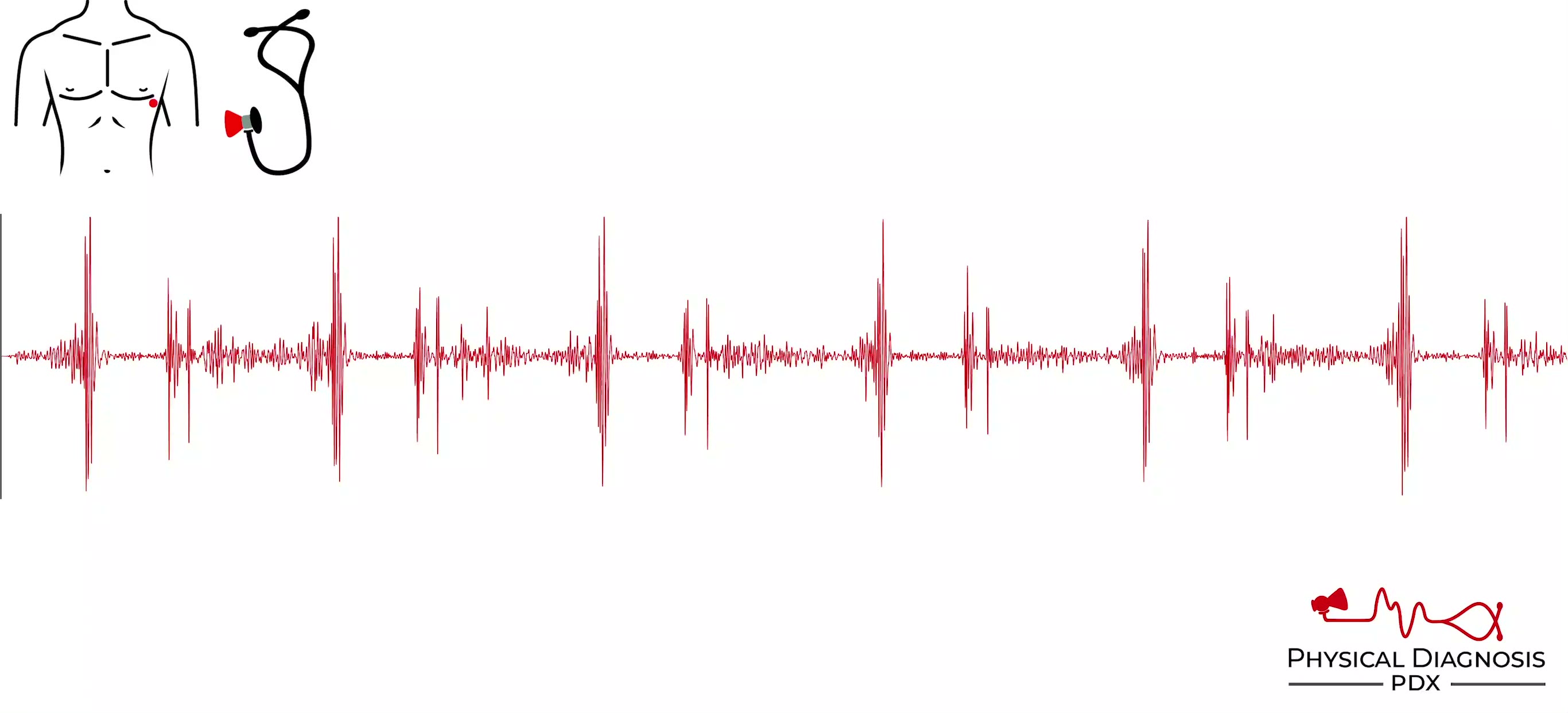
| | | | --- | --- | | **Causes of Loud S1** | | | **Condition** | **Mechanism** | | Short P-R interval (0.08-0.12 second) | Valve wide open from recent atrial contraction | | Premature beats, tachycardia | Valves wide op

Pemberton’s Sign
2 patients · 2 clips
Pemberton’s sign describes the onset of facial plethora after raising the arms above the head, and is found in conditions such as superior vena cava syndrome and large goiter. Card Tutorial Patient 1: This is a middle-ag
Flow Murmur
3 patients · 5 clips
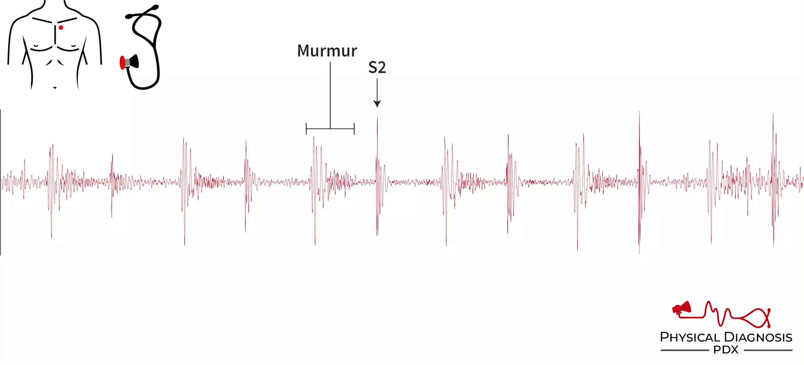
- Flow murmurs may also be referred to as innocent, physiologic, benign, and systolic flow related. - They are typically systolic, short, soft (usually <3/6), crescendo-decrescendo in shape (early-peaking), and heard bes
Friedreich’s Sign
4 patients · 6 clips
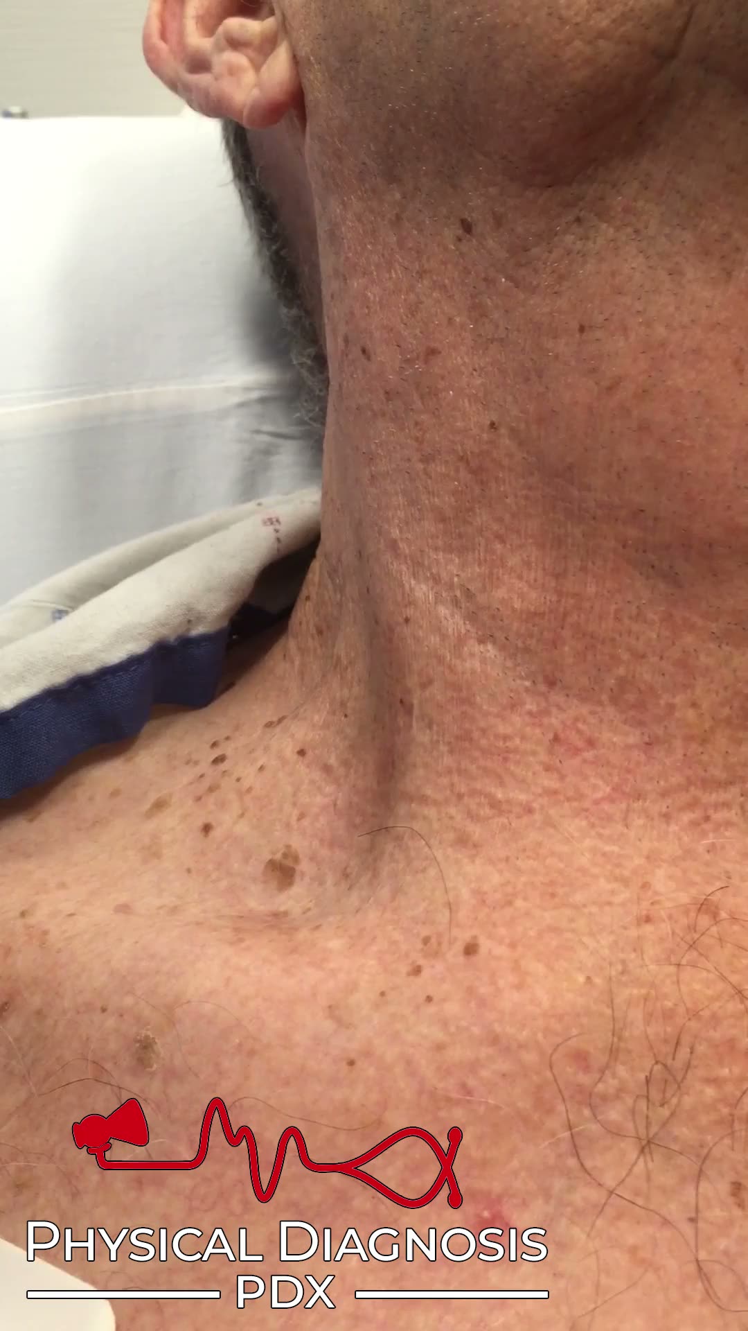
The normal jugular venous waveform contains two descents, x and y. The x descent, which corresponds to the combination of right atrial relaxation and depression of the atrial floor during ventricular contraction, is norm
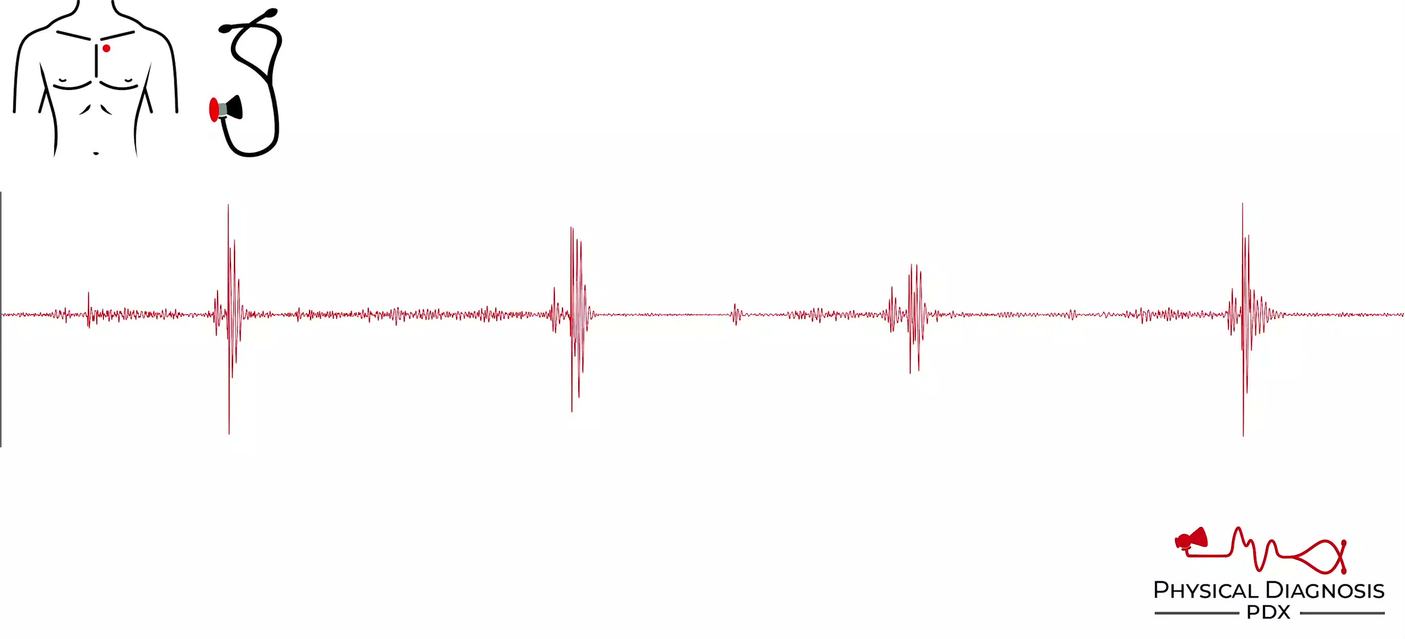
Loud S2
1 patients · 2 clips
The common causes of a loud S2 include high systemic (A2) or pulmonary (P2) pressures and closure of a prosthetic valve (usually in the aortic position). If the S2 is split, try to decipher which component is loudest. A2
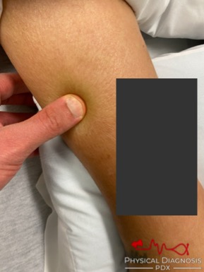
Peripheral Edema
2 patients · 3 clips
Card Tutorial Patient 1: This patient also has Austin Flint murmur and Aortic Regurgitation. Patient 2: This is a young woman with decompensated heart failure and cirrhosis. This patient also has elevated jugular venous
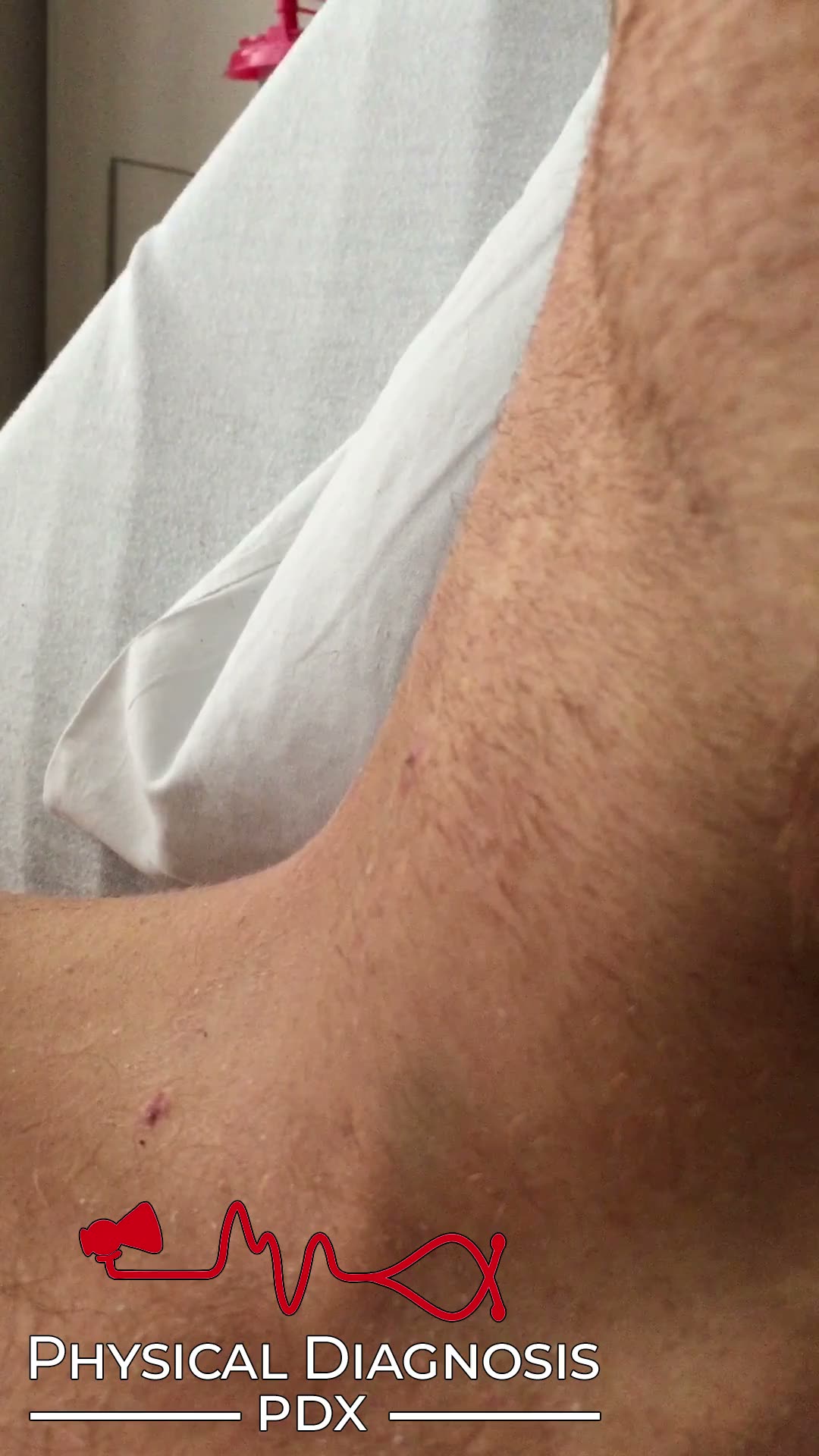
Giant a Wave
7 patients · 9 clips
The a wave of the jugular venous waveform occurs as a result of right atrial contraction. When there is resistance to right ventricular filling, right atrial contraction generates increased pressure, resulting in giant a
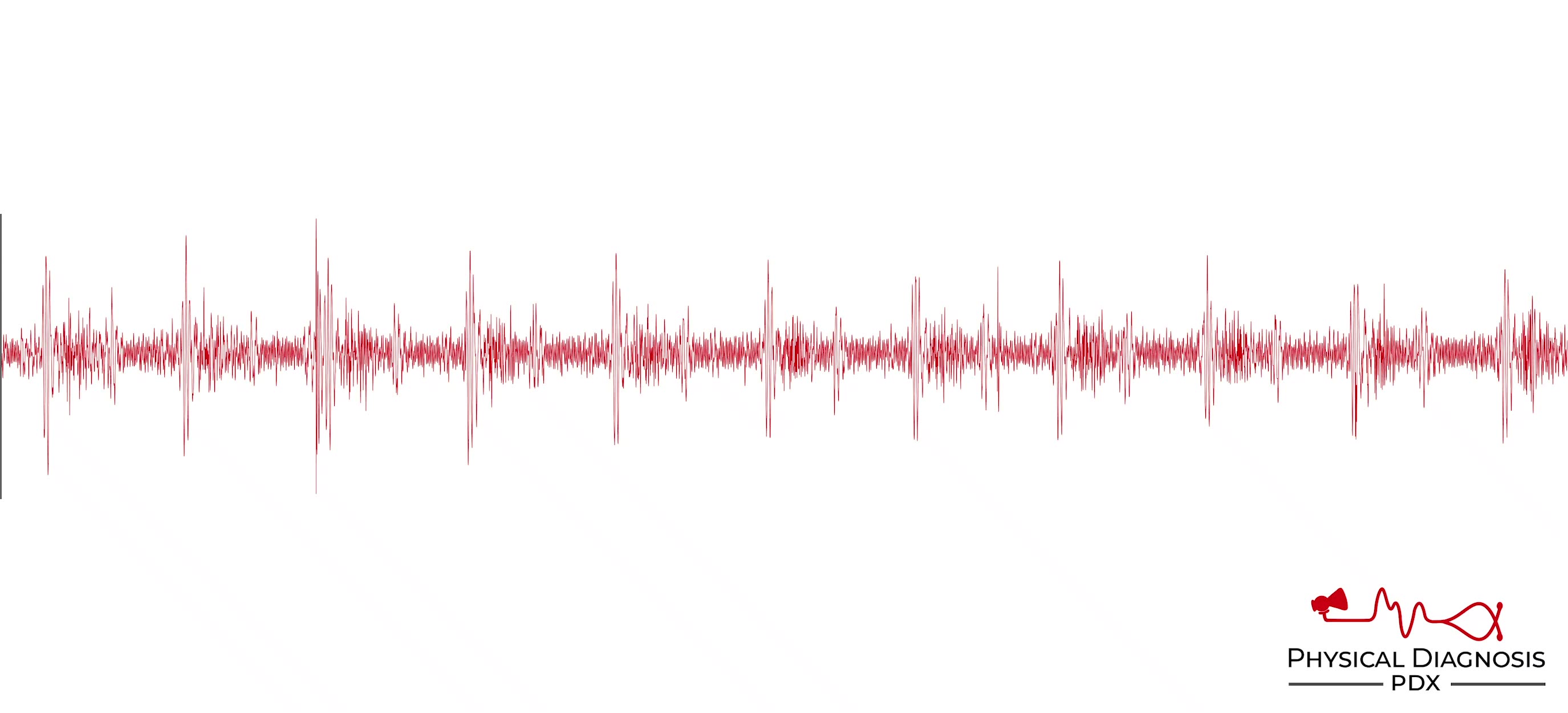
Heart Murmurs
1 patients · 8 clips
Clinical finding with teaching media.
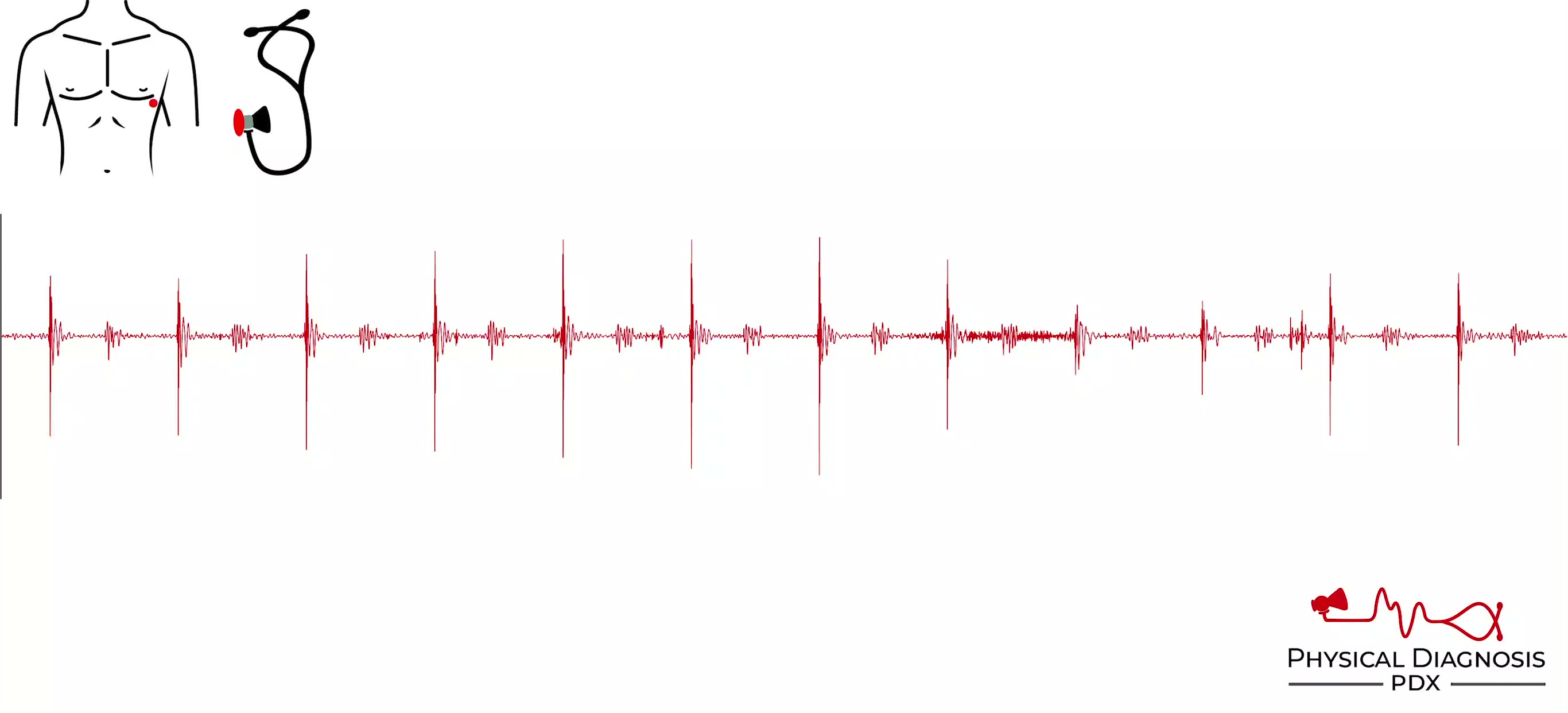
Mechanical S1
1 patients · 2 clips
A mechanical S1 is generated by closure of a prosthetic heart valve (virtually always in the mitral position). It has a “metallic” quality. The presence of a sternotomy scar can provide a clue to the history of valve rep
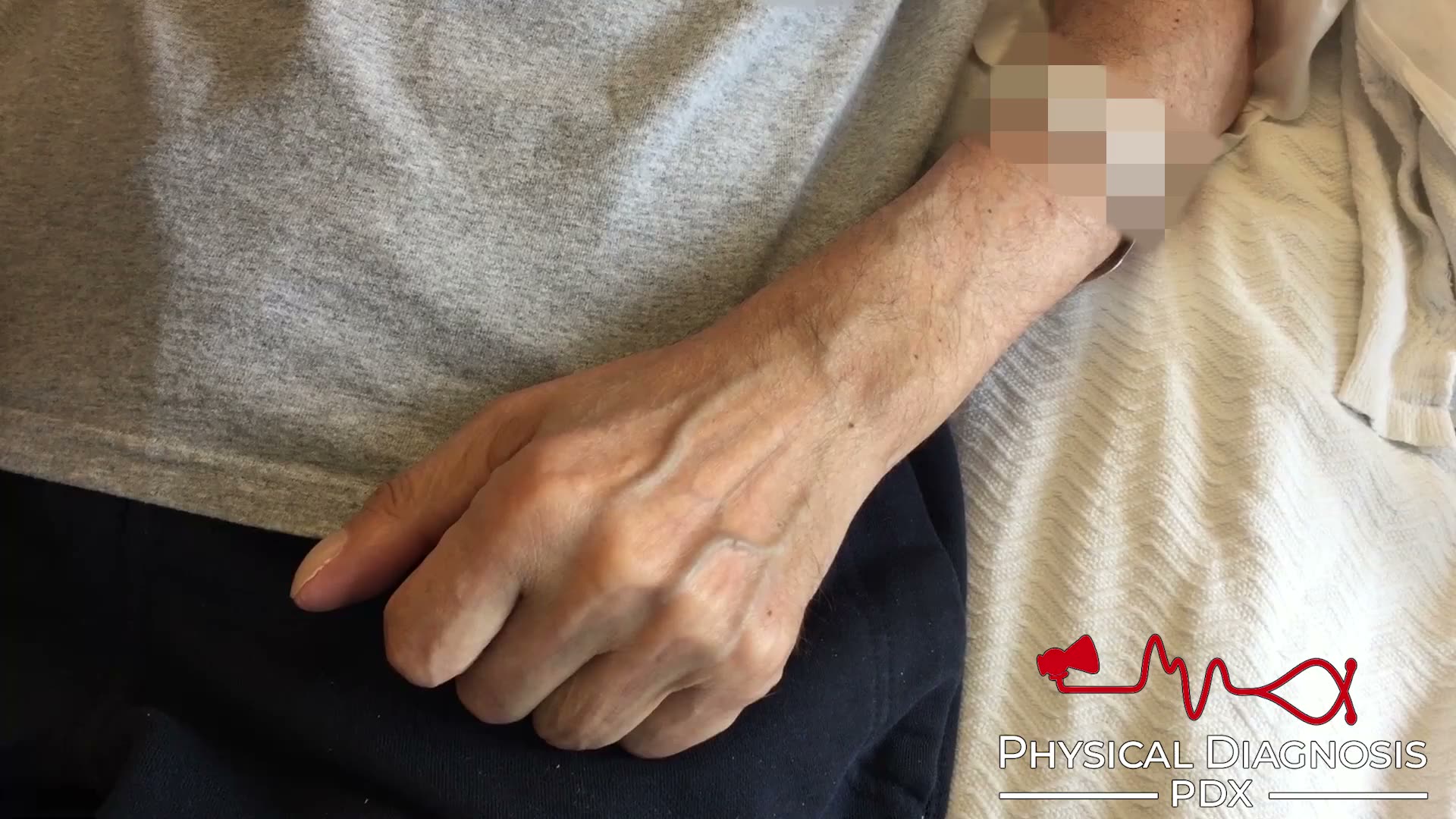
Hand Vein Assessment
3 patients · 3 clips
We have found that the dorsum of the hand can provide fertile ground for estimating central venous pressure. When the internal or external veins are unavailable, try using the hand veins to estimate central venous pressu
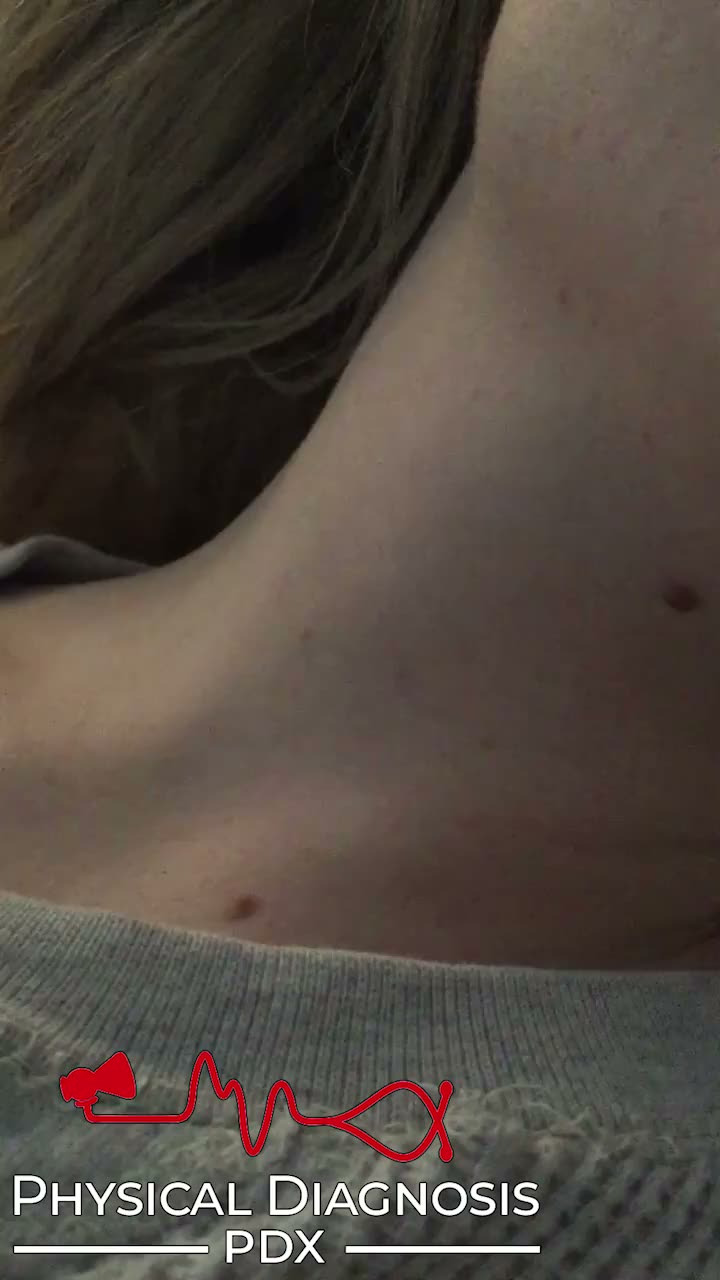
Jugular Venous Pressure via Right Internal Jugular Vein
1 patients · 2 clips
Because of its direct route to the heart, the right internal jugular vein is the preferred site to evaluate the jugular venous pulse. At the bedside, we do not actually visualize the internal jugular vein itself. Rather,
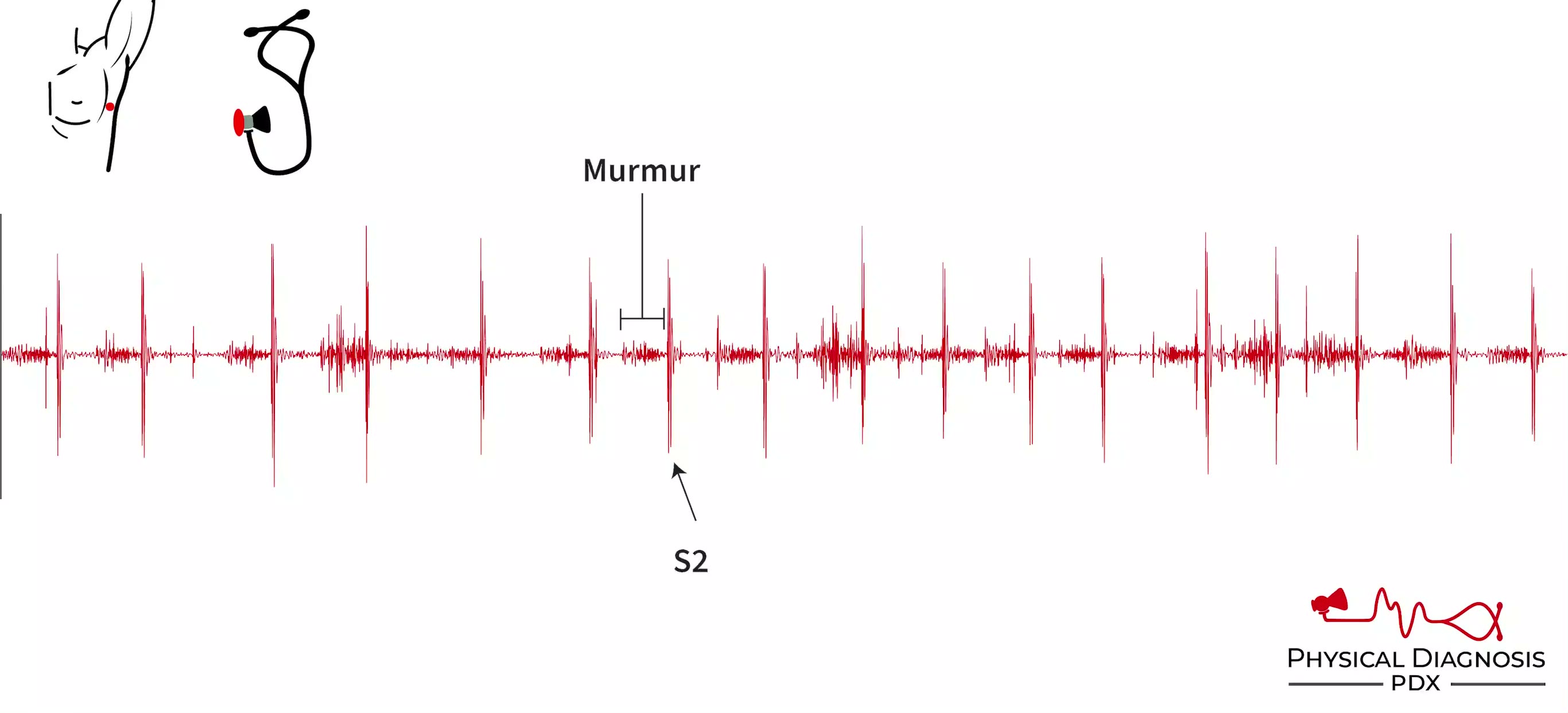
Mitral Regurgitation
16 patients · 39 clips
The MR Murmur is classically holosystolic (from S1 all the way to S2), plateau-shaped. Louder with increased afterload (hand grip or BP cuffs). Loudest at the apex, radiates to the axilla (except in isolated posterior le
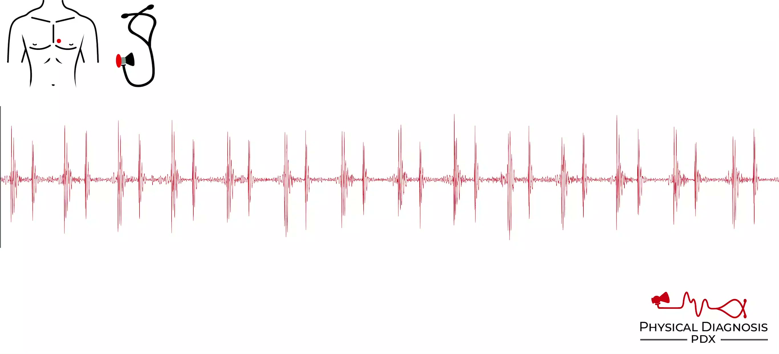
Normal S1
2 patients · 10 clips
Patient 1: This is a young and healthy patient who volunteered to have his heart sounds recorded. **Normal S1** **Annotated** Patient 2: This is a young and healthy medical student who volunteered to have his heart sound

Jugular Venous Pulse in Lung Disease
4 patients · 5 clips
The jugular venous pulse can provide a clue to the presence of lung disease. Decreased intrathoracic pressure during inspiration normally leads to an increase in venous return to the right side of the heart, with an asso
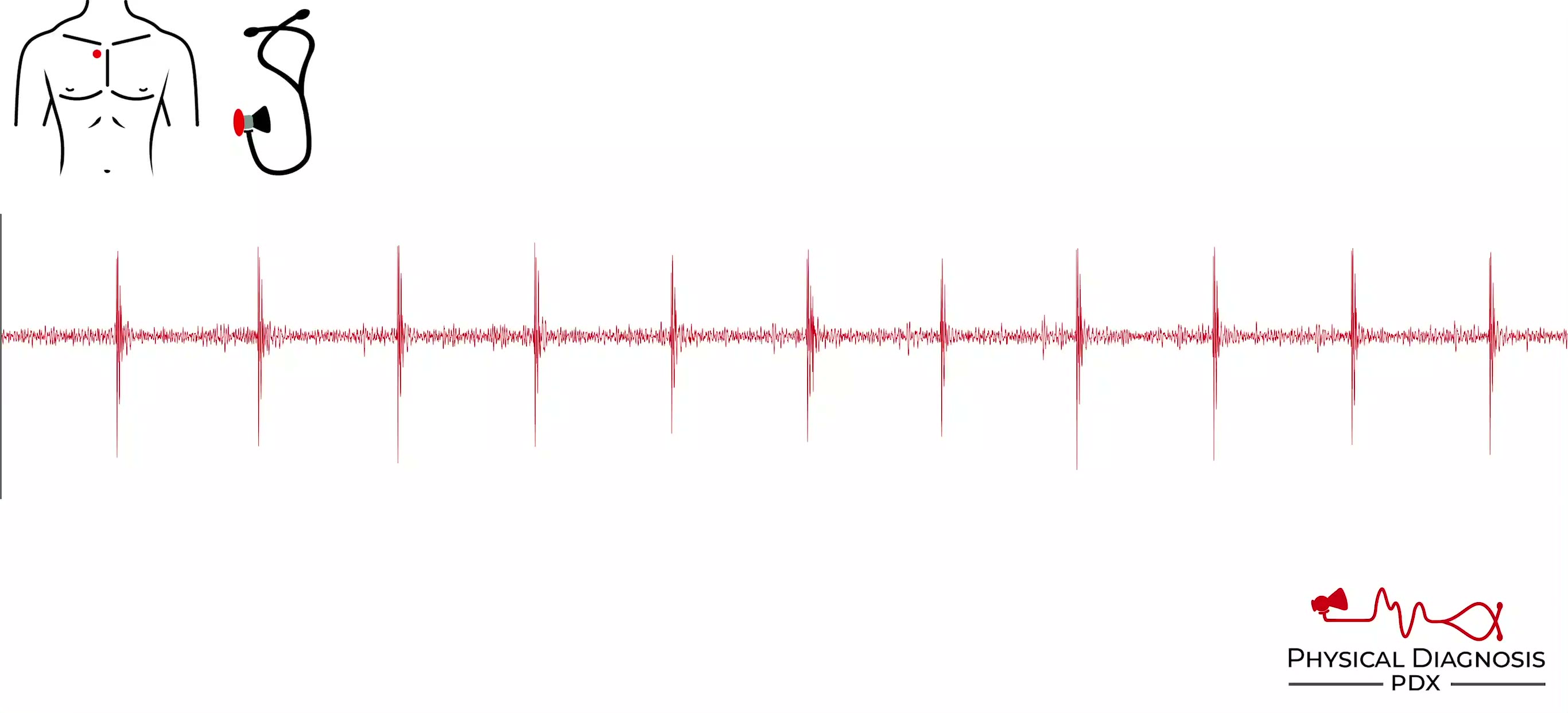
Normal S2
2 patients · 10 clips
Patient 1: This is a young healthy patient who volunteered to have his heart sounds recorded. **Normal S2** **Annotated** Patient 2: This is a young and healthy medical student who volunteered to have his heart sounds re
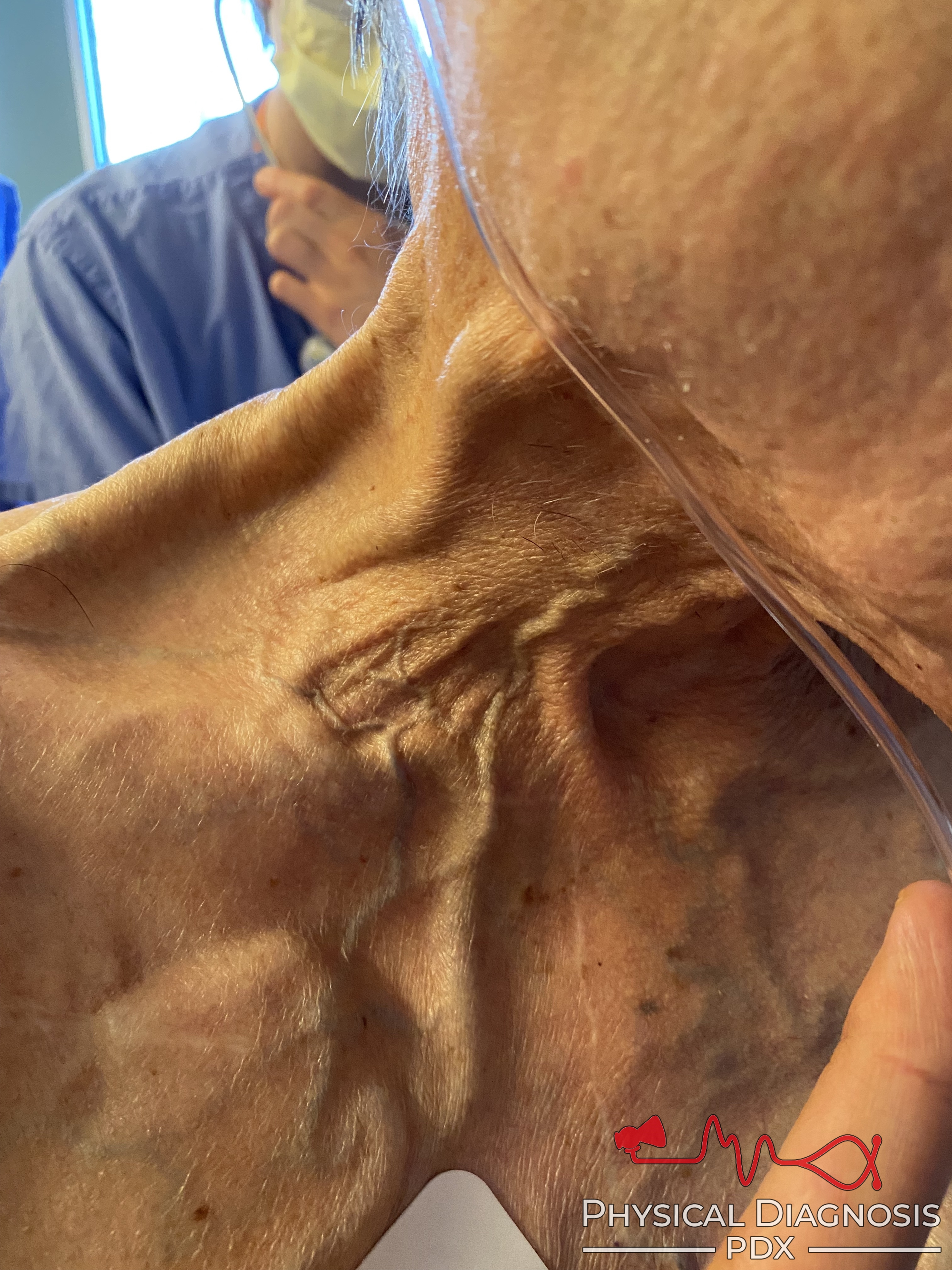
Jugular Venous Pulse via External Jugular Vein
5 patients · 7 clips
When the right and left internal jugular veins fail to provide adequate evaluation of the jugular venous pulse, the external jugular vein can be used to estimate central venous pressure. The external jugular is easily re
Paradoxically Split S2
A paradoxically split S2 describes the presence of a split S2 during expiration only (the opposite of a physiologically split S2). This is caused by any condition that delays aortic valve closure. During expiration, the
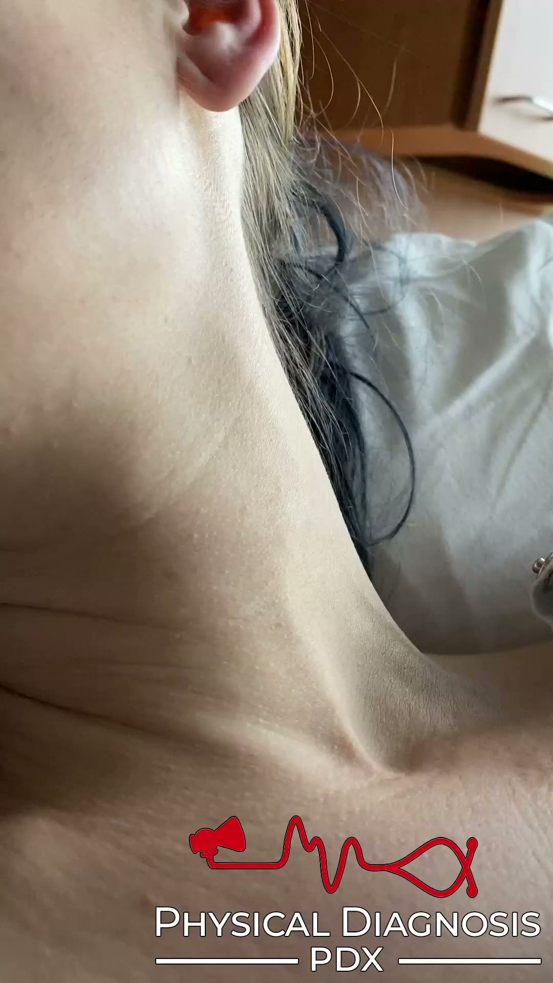
Jugular Venous Pulse via Left Internal Jugular Vein
2 patients · 2 clips
The right internal jugular vein is the preferred site to evaluate the jugular venous pulse. Sometimes it is unavailable because of thrombosis or the presence of a catheter. In other patients, it is simply not well seen o
Pericardial Friction Rub
8 patients · 14 clips
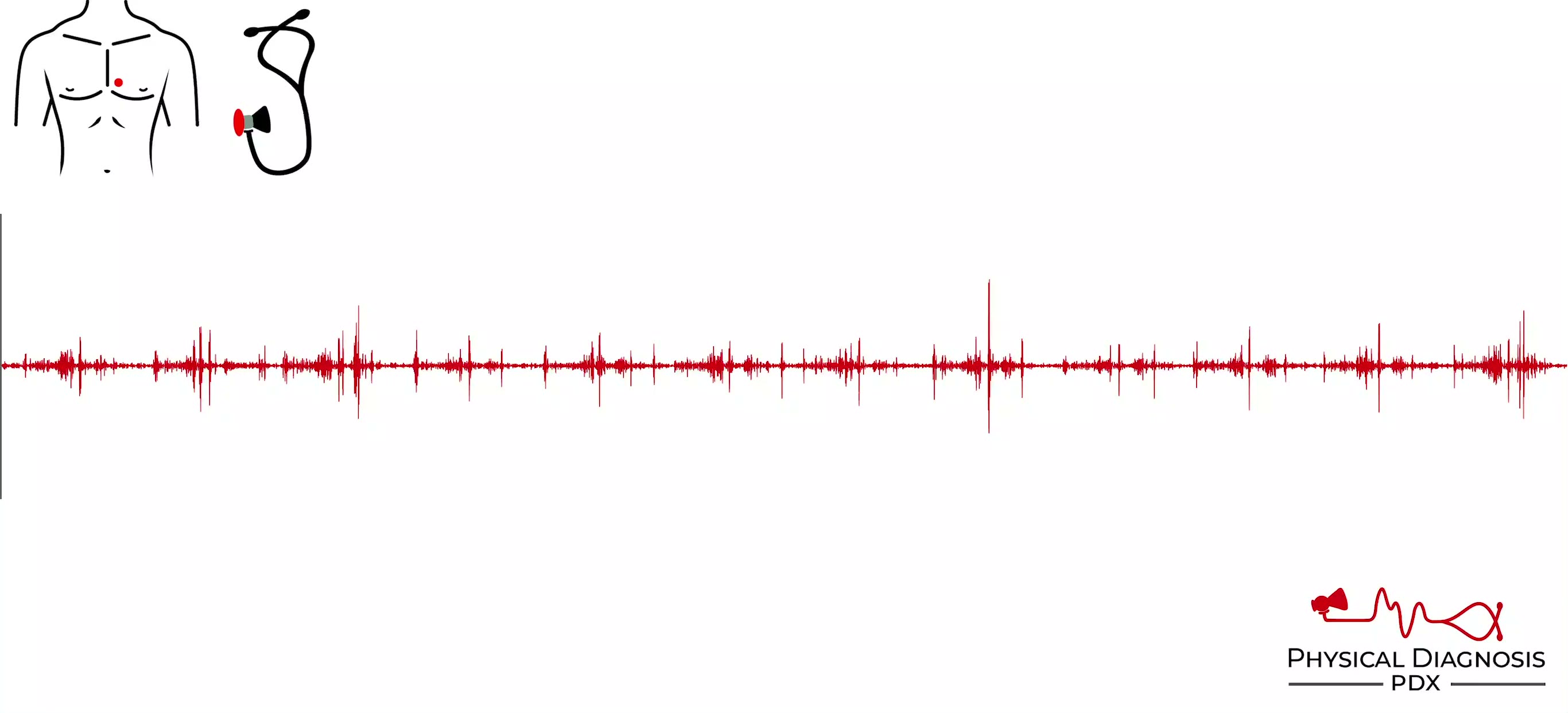
- A pericardial friction rub is the sound that is generated as a result of an inflamed pericardium. - Rubs consist of one to three sounds that may be mistaken for murmurs. However, the quality of the sound(s) is differen
Pulmonic Regurgitation
4 patients · 8 clips
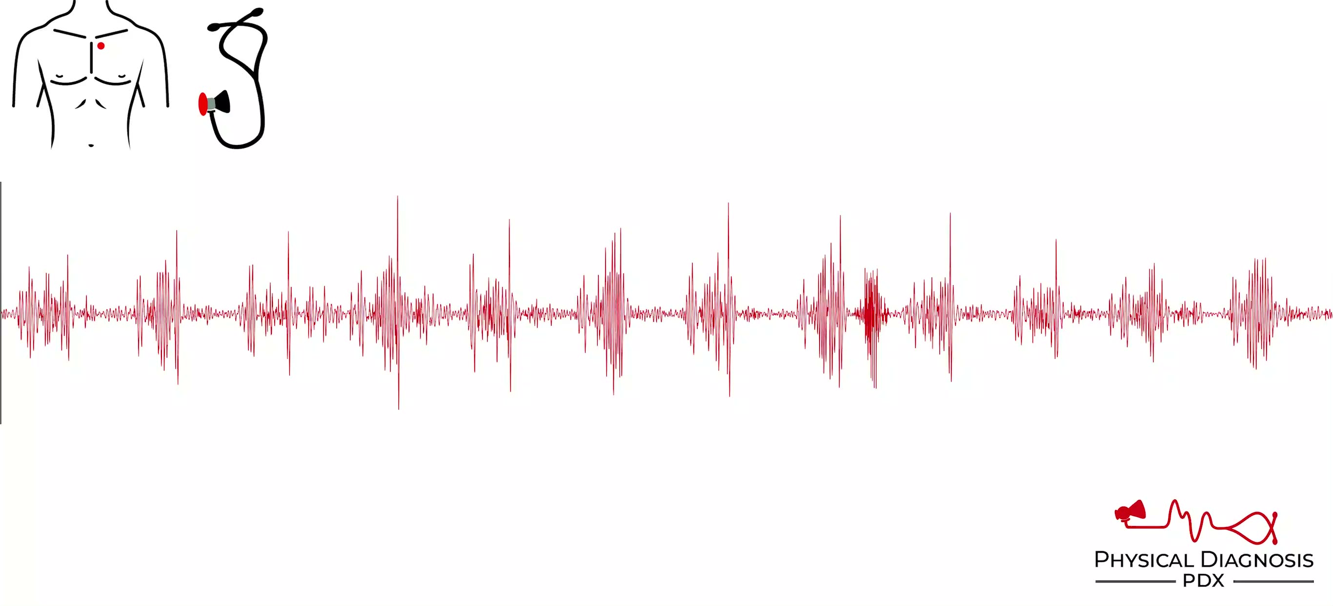
- There are a variety of causes of pulmonic regurgitation, including pulmonary hypertension (so-called Graham-Steell murmur), infective endocarditis, and congenital abnormalities of the cusps (eg, bicuspid valve). - The
Jugular Venous Waveform in Atrial Fibrillation
2 patients · 3 clips
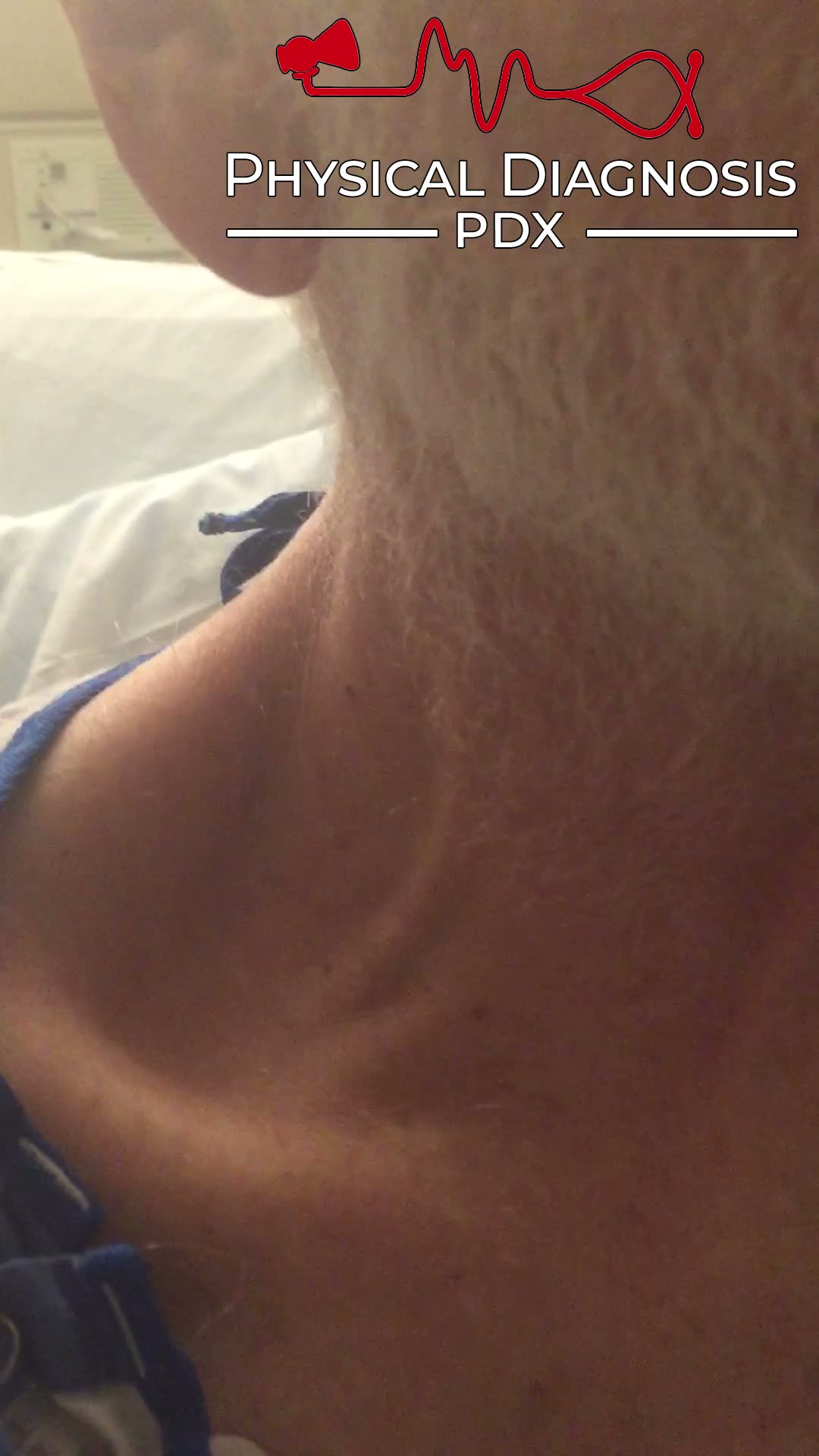
The jugular venous pulse can provide a clue to the diagnosis of atrial fibrillation. The a wave of the jugular venous waveform occurs as a result of right atrial contraction. Because atrial fibrillation does not allow fo
Pericardial Knock
3 patients · 13 clips
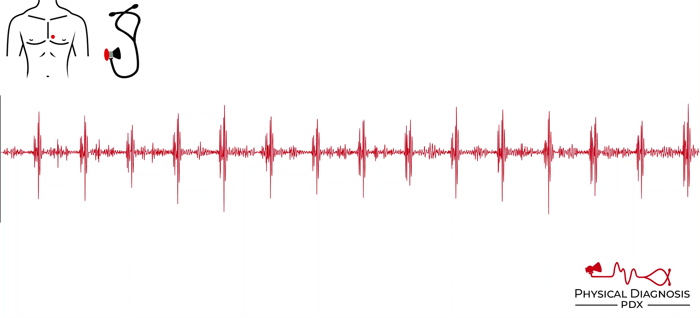
The pericardial knock is a loud, high pitched sound in early diastole that occurs in patients with constrictive pericarditis. It is the result of rapid deceleration of blood due to the inelastic pericardium. It occurs we
Pulmonic Stenosis
2 patients · 4 clips
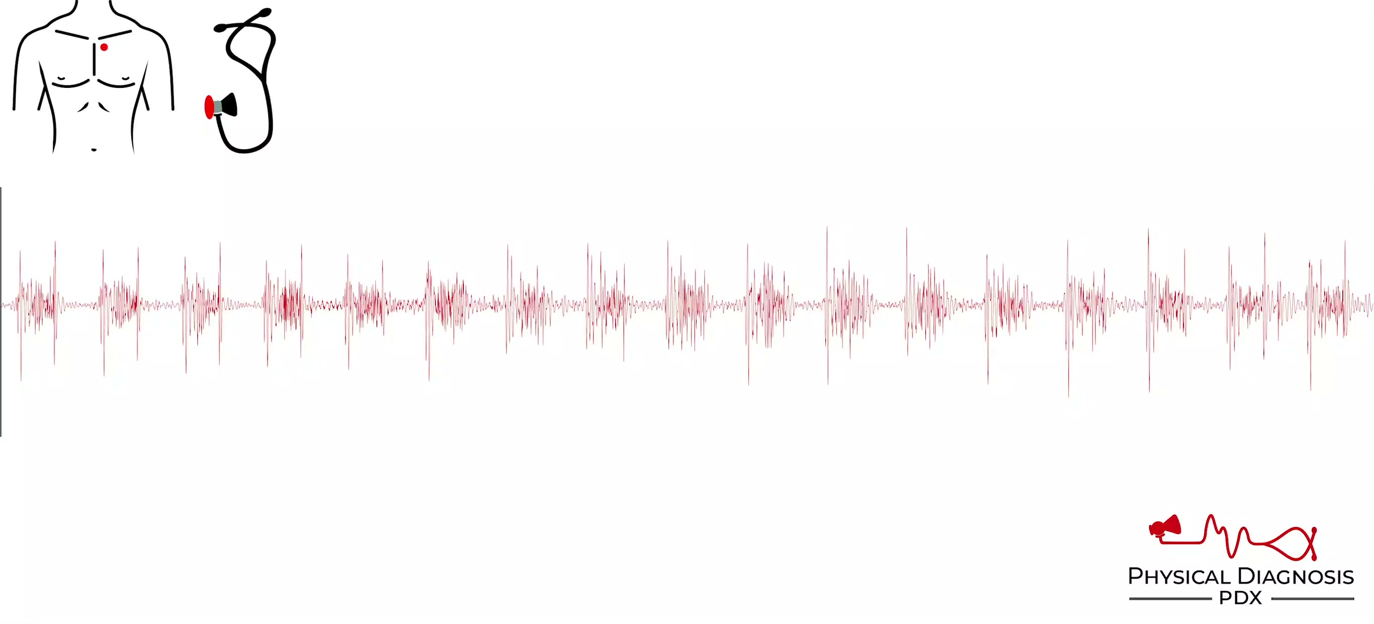
- Pulmonic stenosis is virtually always congenital. However, it may occur as a result of acquired conditions such as rheumatic disease and carcinoid syndrome. - The systolic murmur of pulmonic stenosis is best heard over
JVP Forearm Assessment
1 patients · 1 clips
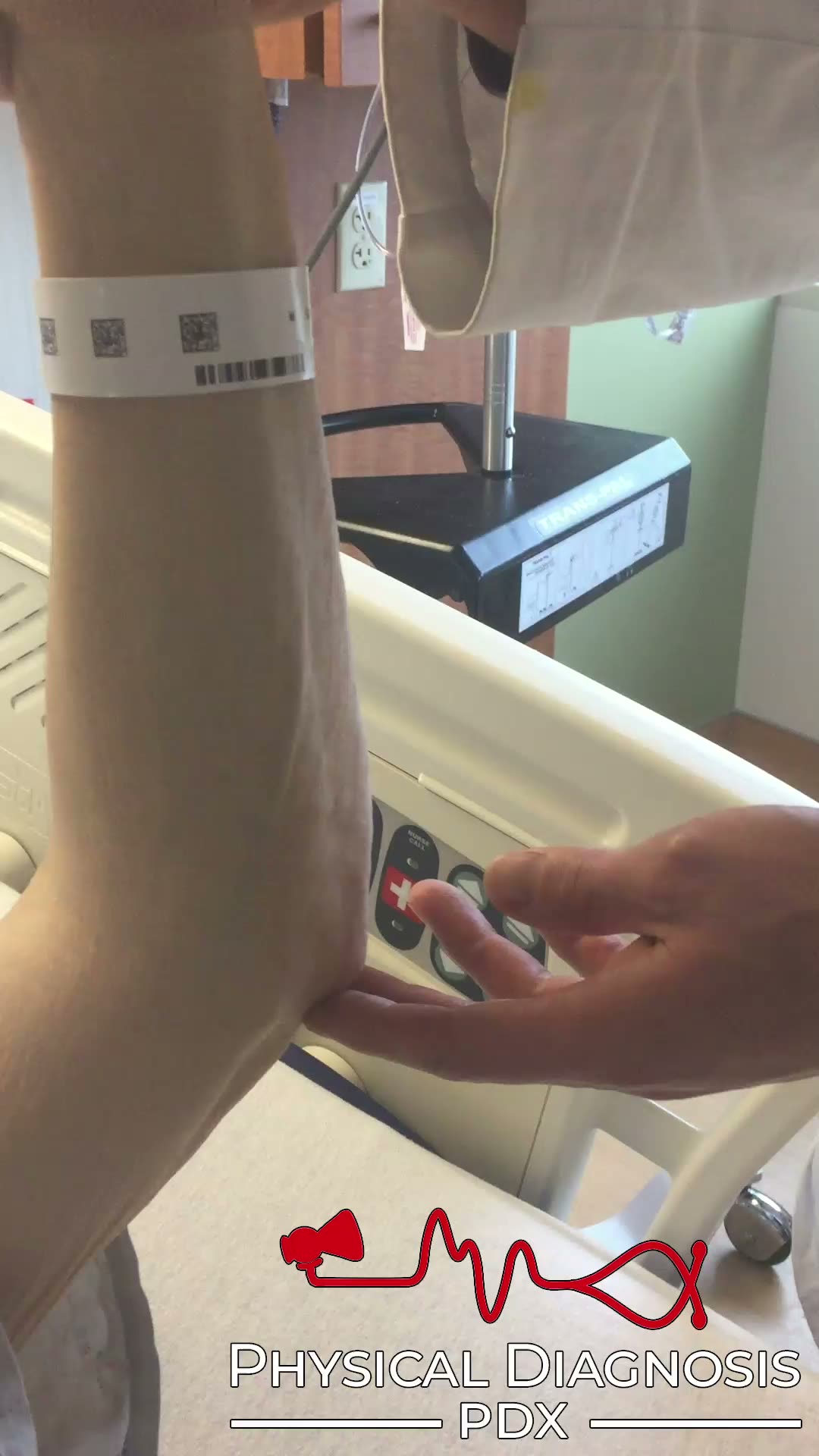
Pulmonology Tutorial Patient 1: This patient has
Systolic Murmur
1 patients · 1 clips
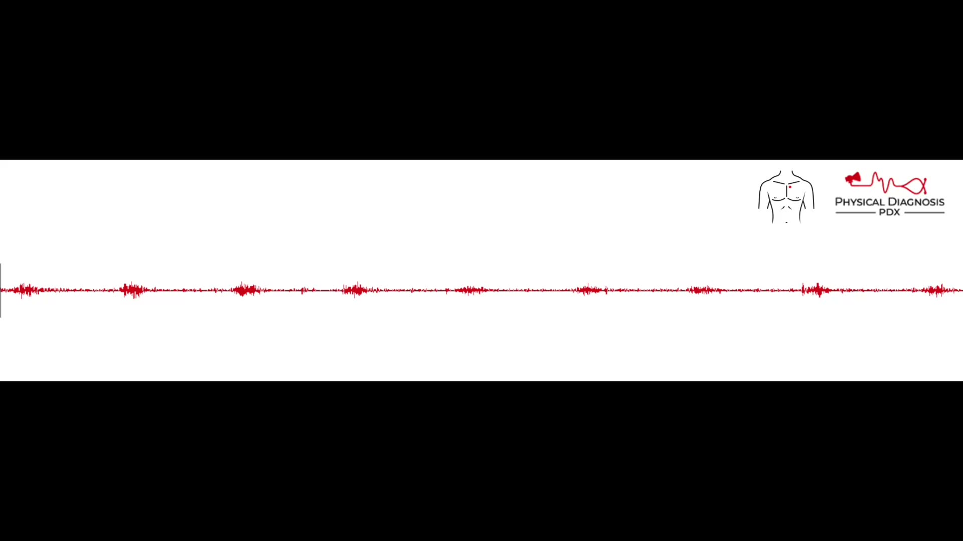
Patient 1: 43 y/o man with a history of total anomalous pulmonary venous drainage s/p corrective surgery at age 3, found to have a systolic murmur over the LUSB, which is brought about by lifting the left arm above the l
Kussmaul’s Sign
12 patients · 14 clips
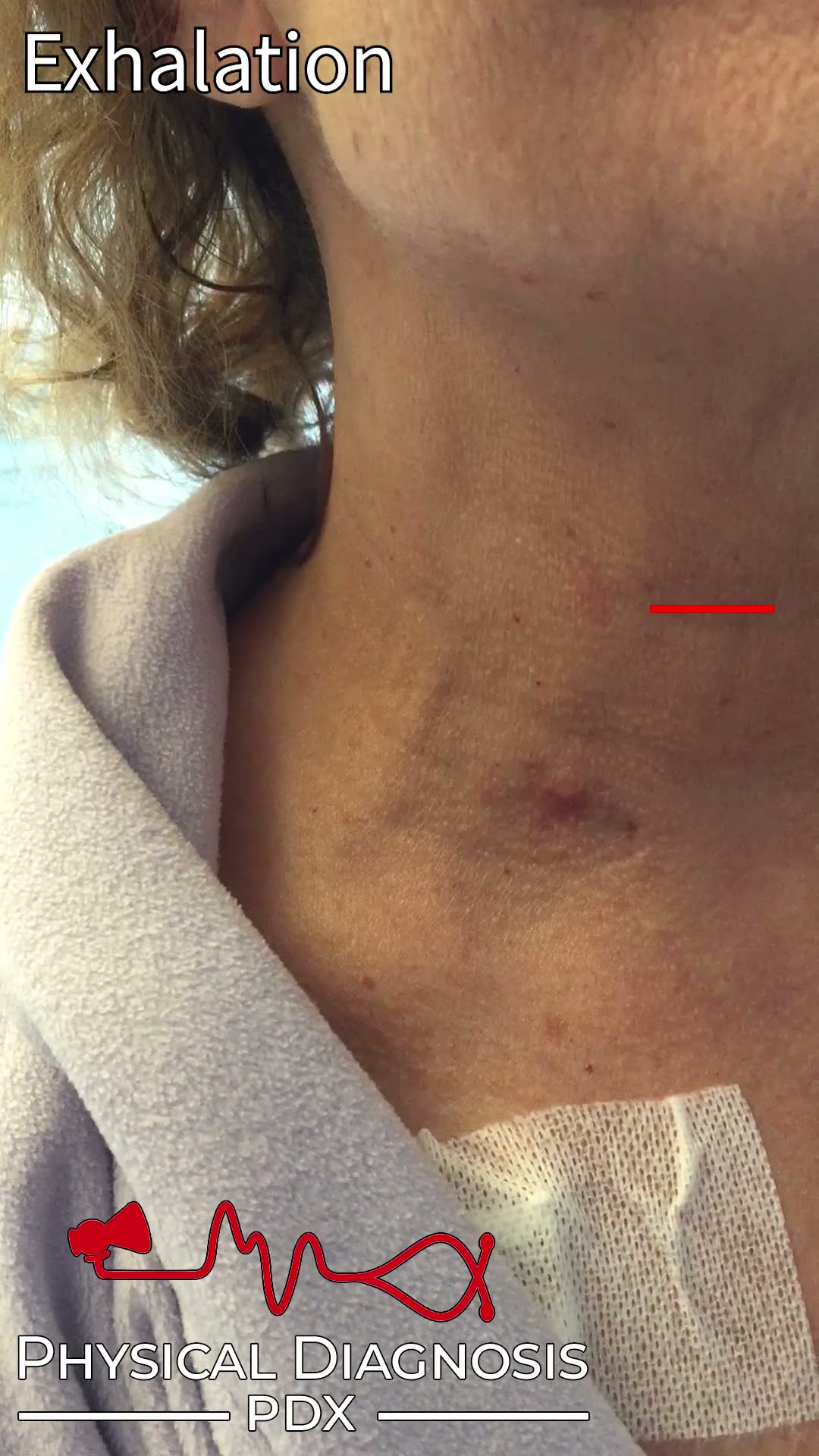
Kussmaul’s sign is defined as a paradoxical rise in jugular venous pressure during inspiration. Decreased intrathoracic pressure during inspiration normally leads to an increase in venous return to the right side of the
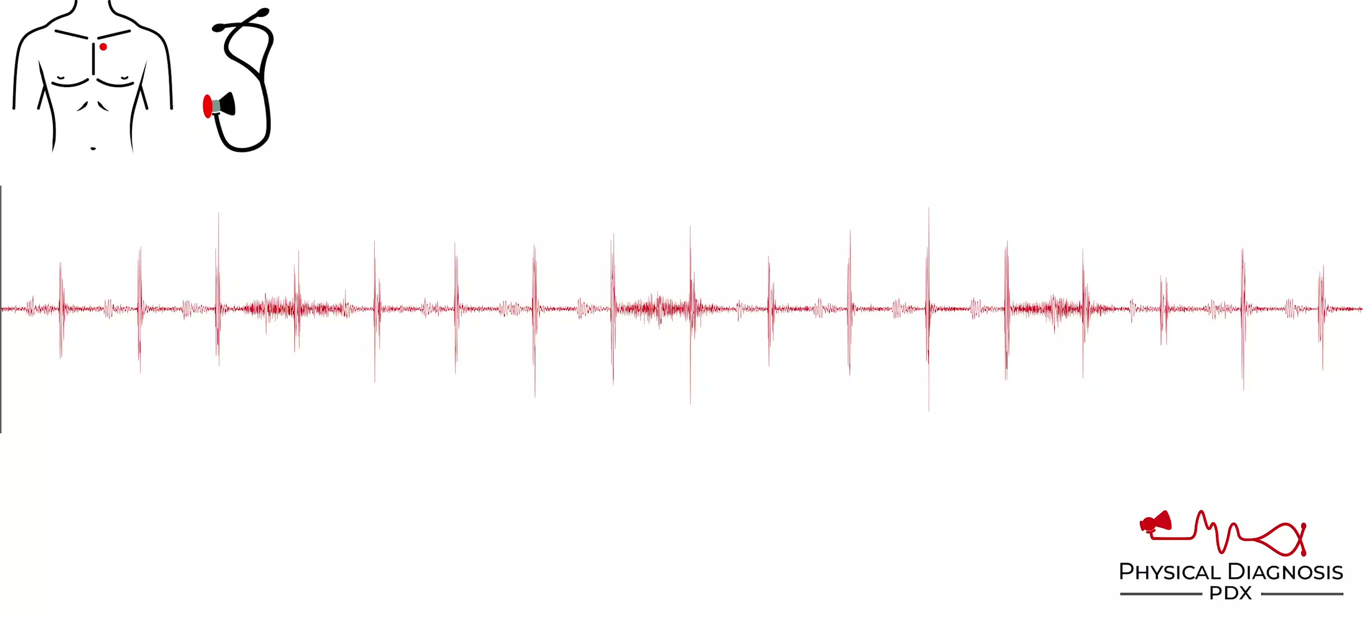
Physiologically Split S2
6 patients · 11 clips
Normally the higher pressure left -sided valves close first (mitral before tricuspid, aortic before pulmonic). This discordance is exaggerated by inspiration, allowing the human ear to perceive two distinct sounds. Durin
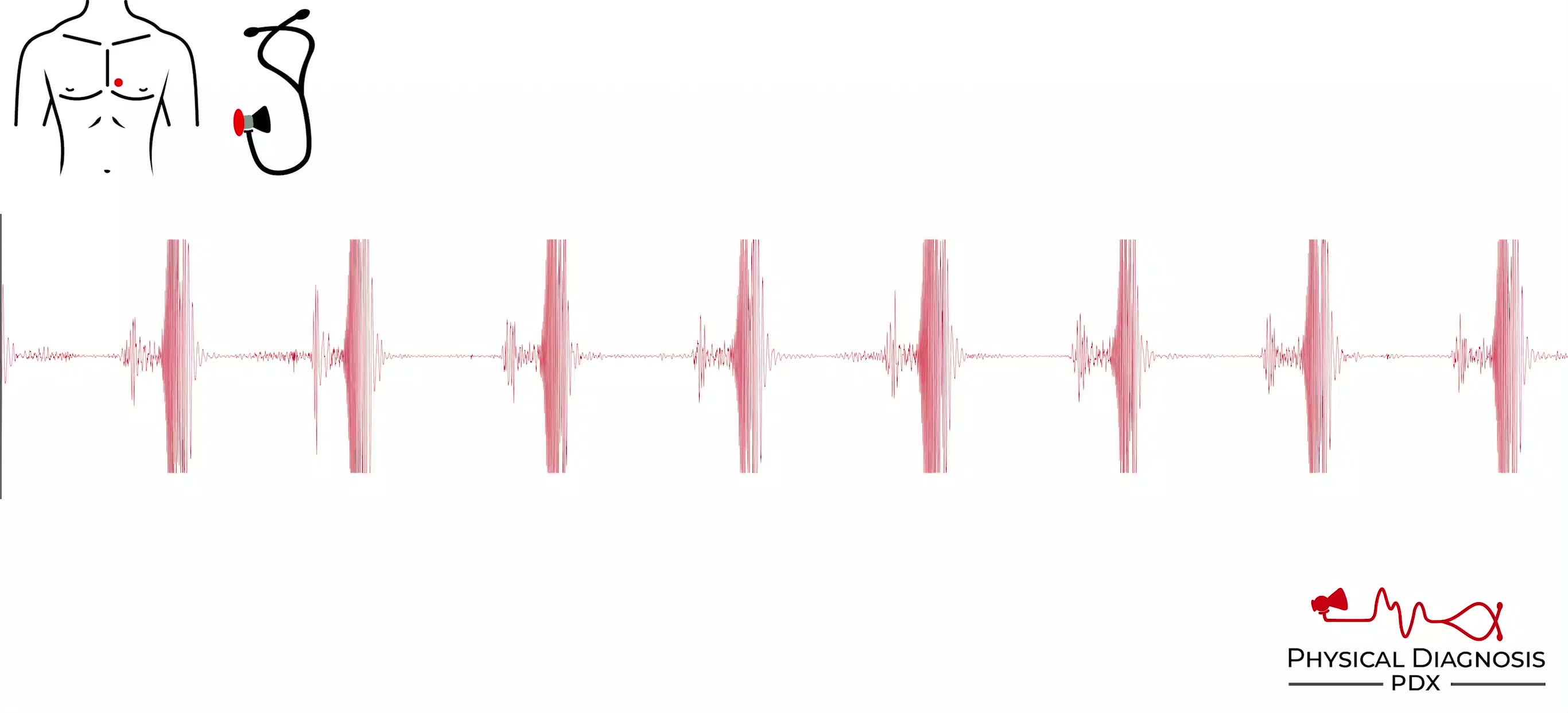
Tricuspid Regurgitation
9 patients · 18 clips
- Tricuspid regurgitation is a fairly common lesion in the 21st century, likely due to the large number of patients who are surviving longer after bouts of endocarditis, pulmonary hypertension, severe left heart disease,
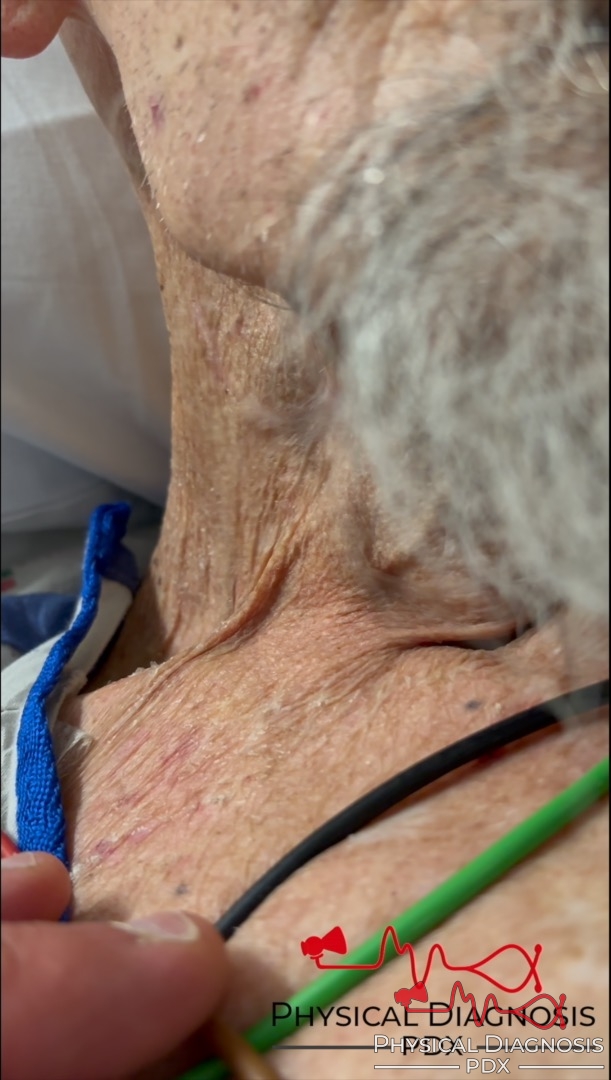
Lancisi’s Sign
12 patients · 18 clips
In the setting of severe tricuspid regurgitation, retrograde blood flow into the right atrium during ventricular systole results in loss of the x descent, creating a fused cv wave that appears as a large pulsation within
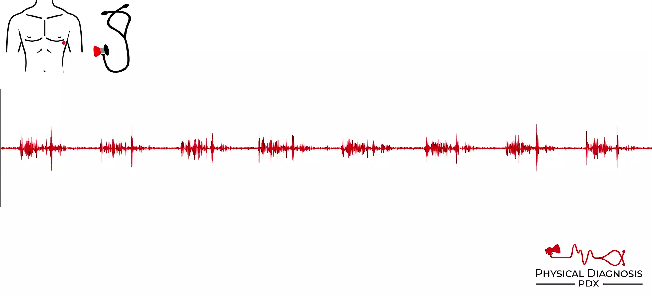
S3 Gallop
11 patients · 21 clips
The S3 occurs during the rapid filling phase of early diastole (the y descent of the jugular venous waveform), 0.14-0.22 seconds after S2 (MUCH farther from S2 than a widely split S2). Unlike the split S2, the S3 is best
Tricuspid Stenosis
5 patients · 12 clips
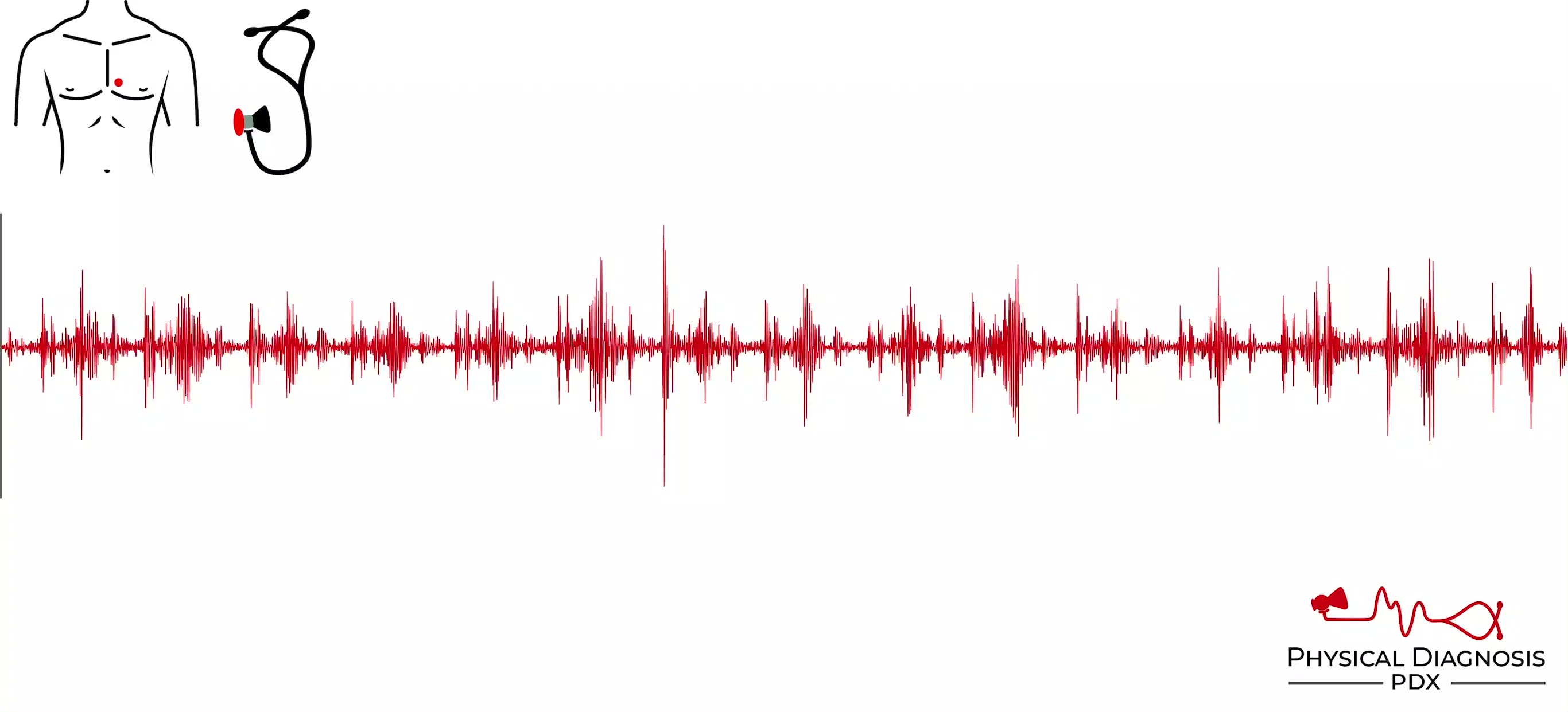
- Tricuspid stenosis is a diastolic murmur similar in quality to mitral stenosis, but is best heard over the left lower sternal border, or between the left lower sternal border and the apex. It also becomes louder with i
Low Jugular Venous Pressure
1 patients · 1 clips
Patient 1: This is a middle-age patient with cardiac sarcoidosis who was admitted to the hospital with decompensated heart failure and subsequently developed acute kidney injury after overly aggressive diuresis. The JVP
S4 Gallop
11 patients · 33 clips
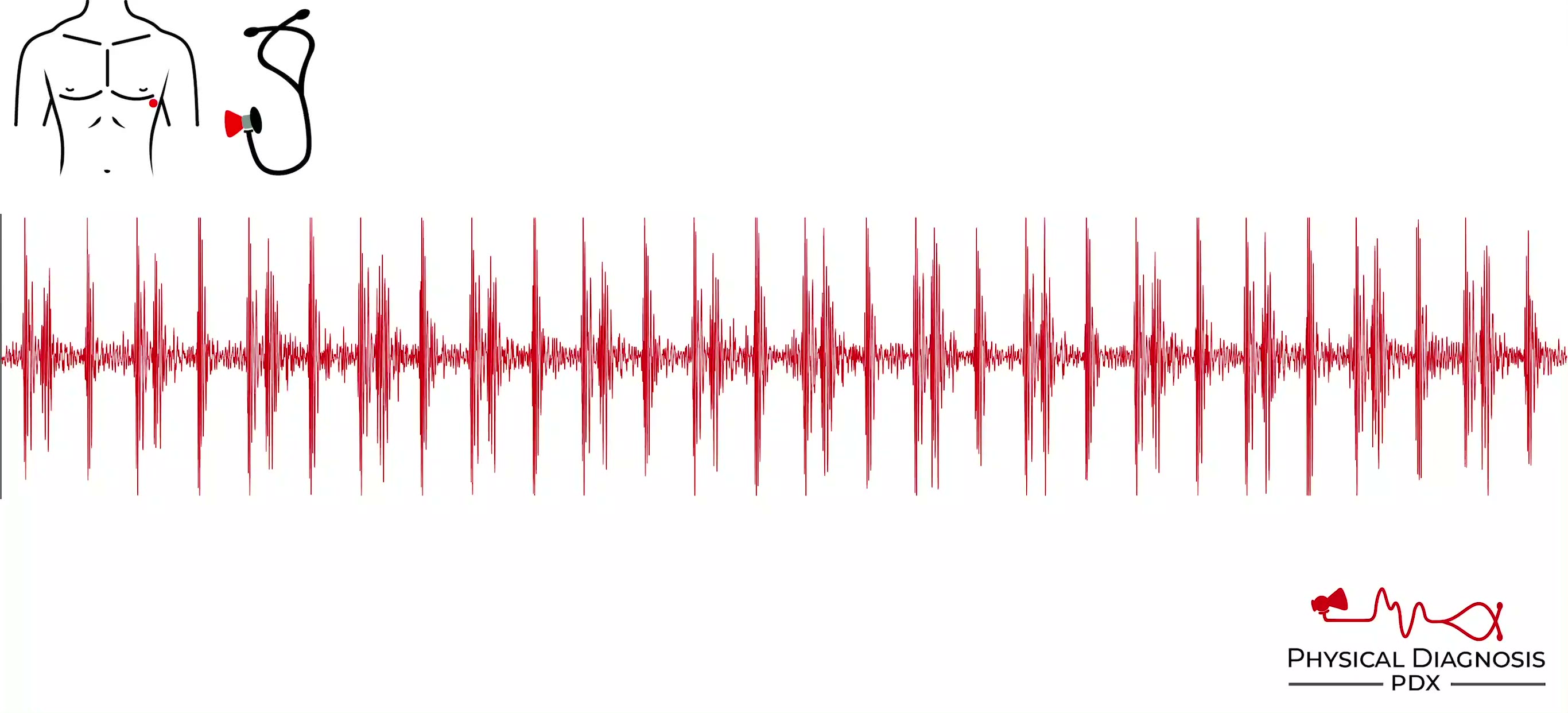
The S4 occurs in late diastole, just before S1, coinciding with atrial contraction (the a wave in the jugular venous waveform). It is generally present when the heart is so stiff that it is reliant upon a significant amo
Venous Hum
2 patients · 6 clips
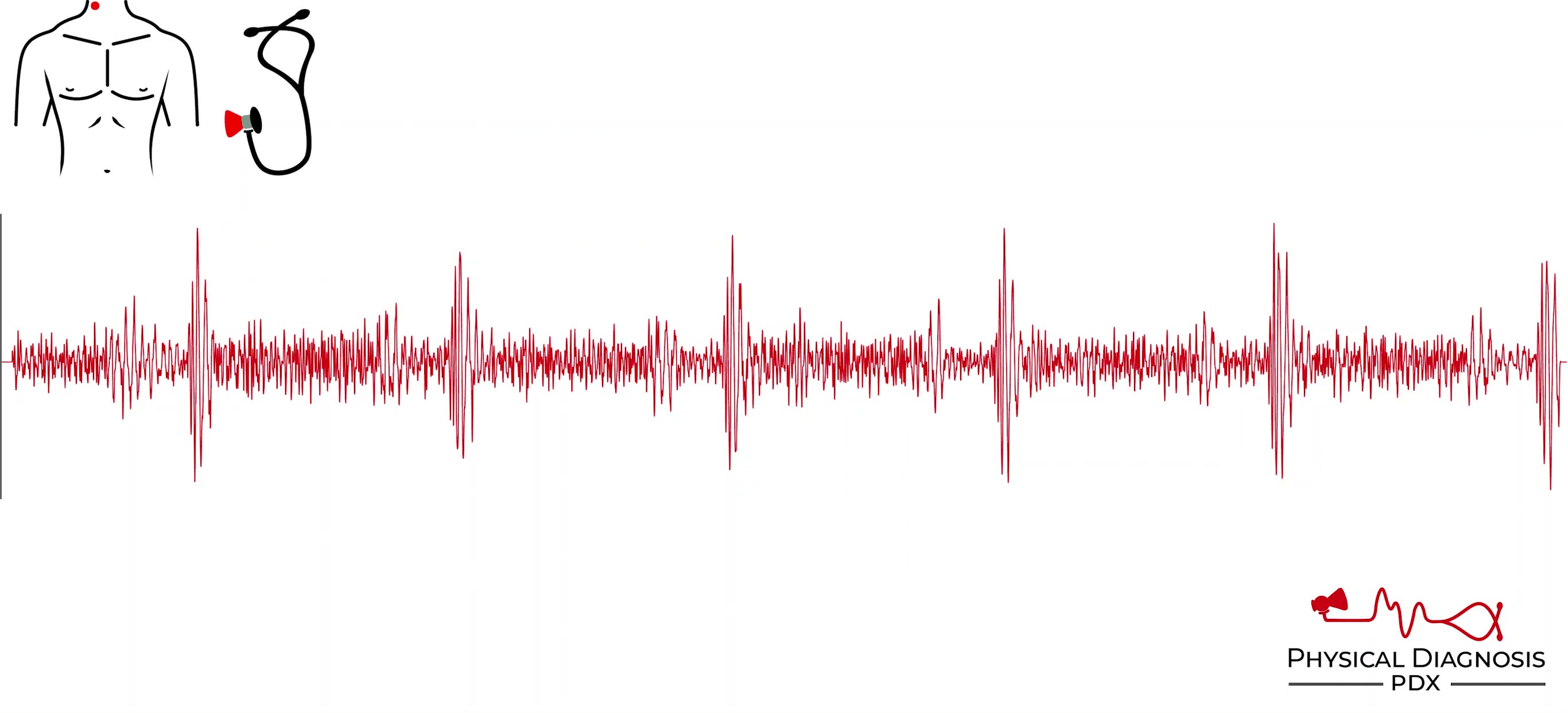
- Often heard in children. - Etiology is thought to be compression of the internal jugular vein by the transverse process of the atlas. - The sound is continuous, loudest in diastole. - Best heard in the neck, in the ant
Normal Jugular Venous Pressure
4 patients · 7 clips
The jugular venous pulse can be used to measure right atrial pressure. Normal right atrial pressure is less than 8 cm H2O (via exam) or less than 6 mm Hg (via direct measurement). The ability to recognize normal right at
Soft S1
3 patients
The common causes of a soft S1 include weak ventricular contractions, long PR interval, emphysema, obesity, and pericardial effusion. Card Tutorial Patient 1: This patient has Patient 2: This patient has Patient 3: This
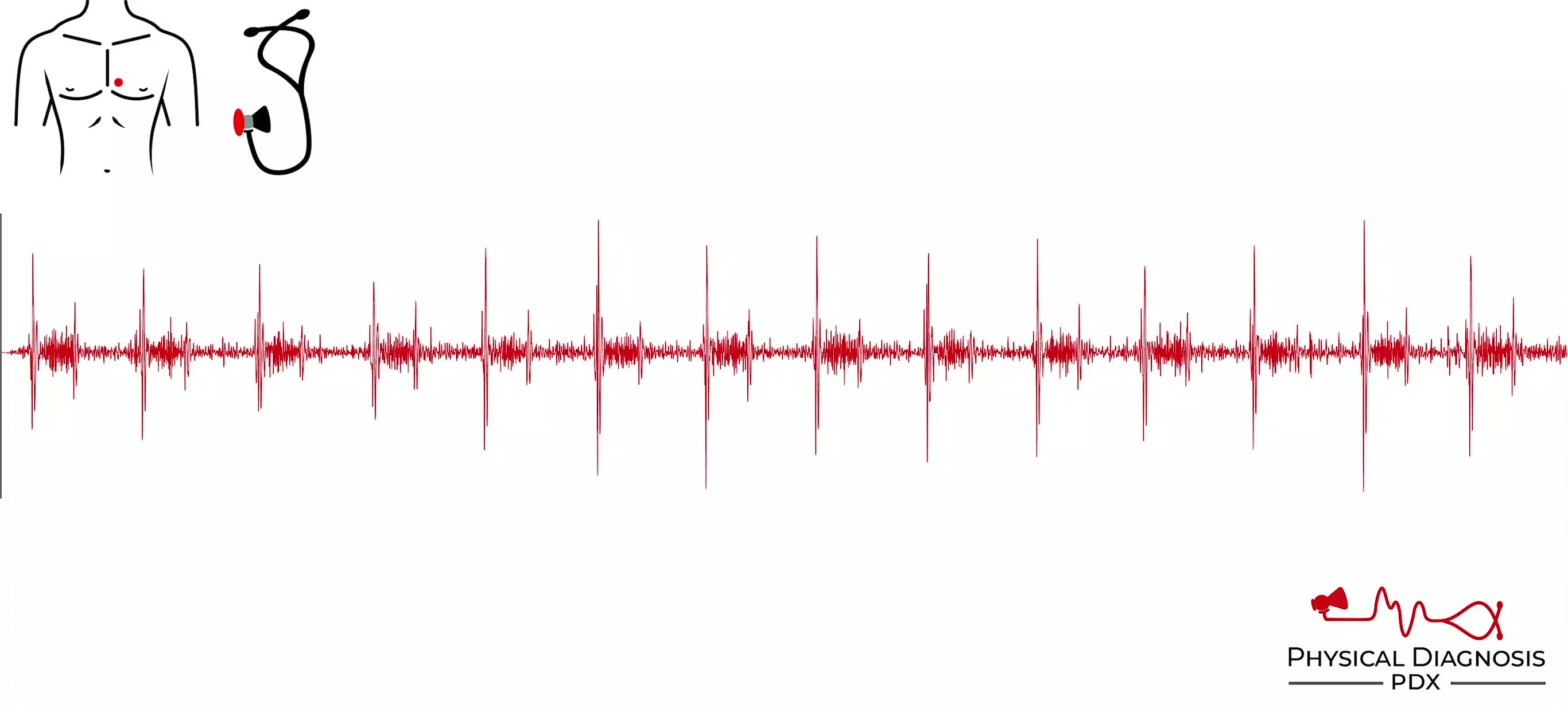
Ventricular Septal Defect
2 patients · 4 clips
The VSD murmur is classically a holosystolic murmur similar to mitral regurgitation but located along the lower left sternal border, often with a palpable thrill. Flow is left to right (as pressure is higher on the left)

Normal Jugular Venous Waveform
5 patients · 9 clips
The normal jugular venous pulse is best evaluated via the right internal jugular vein, and is characterized by three peaks and two troughs. The first peak, called the a wave, results from atrial contraction during late d
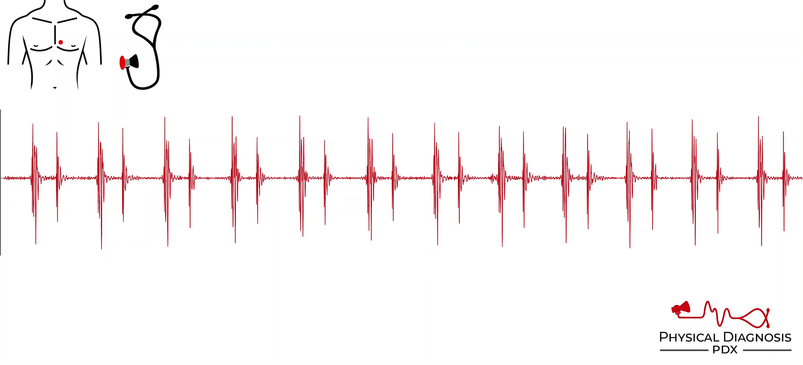
Split S1
4 patients · 8 clips
S1 is generated by closure of the mitral and tricuspid valves. These paired left- and right-sided valves do not close at exactly the same time, but the human ear cannot distinguish sounds that are 0.02-0.03 seconds apart
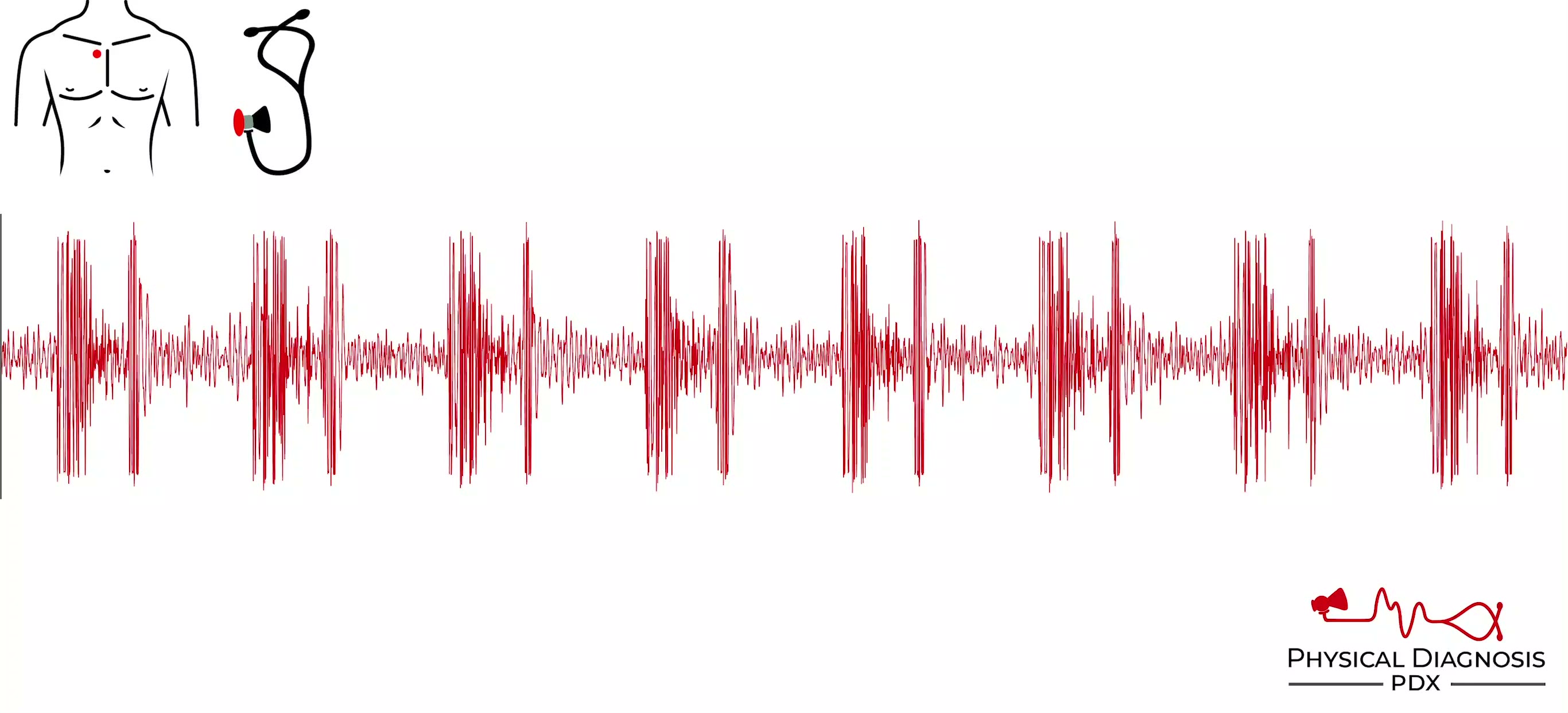
Starr-Edwards Prosthetic Valve
2 patients · 6 clips
- The old “ball-in-cage” prosthetic valve was designed by Albert Starr and Lowell Edwards and implanted into a patient for the first time in 1960. - The design of the prosthetic valve is unique in that increased pressure
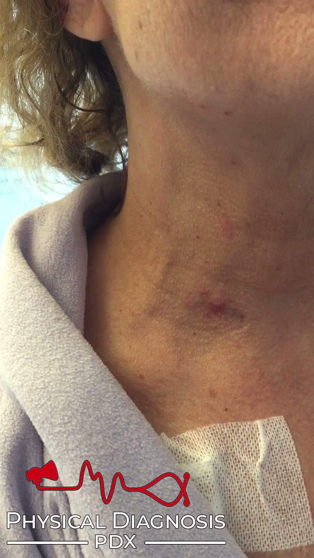
W Sign
1 patients · 2 clips
The normal jugular venous waveform contains two descents, x and y. Under normal conditions, the x descent is more prominent than the y descent. In constrictive pericarditis, the unyielding pericardium causes elevated atr
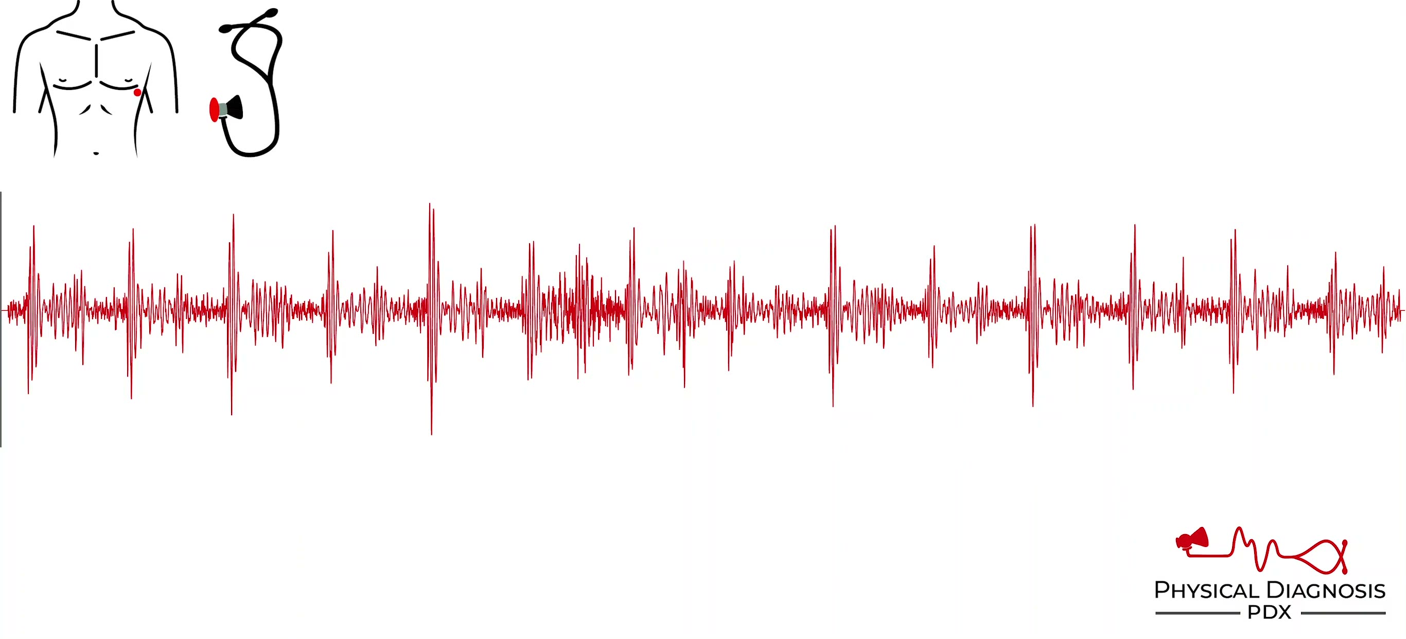
Summation Gallop
3 patients · 4 clips
Patient 1: This is a young woman with new systolic heart failure related to drug use. There is a gallop and because of tachycardia, it is difficult to determine whether it’s an S3 or an S4. It could be a summation gallop
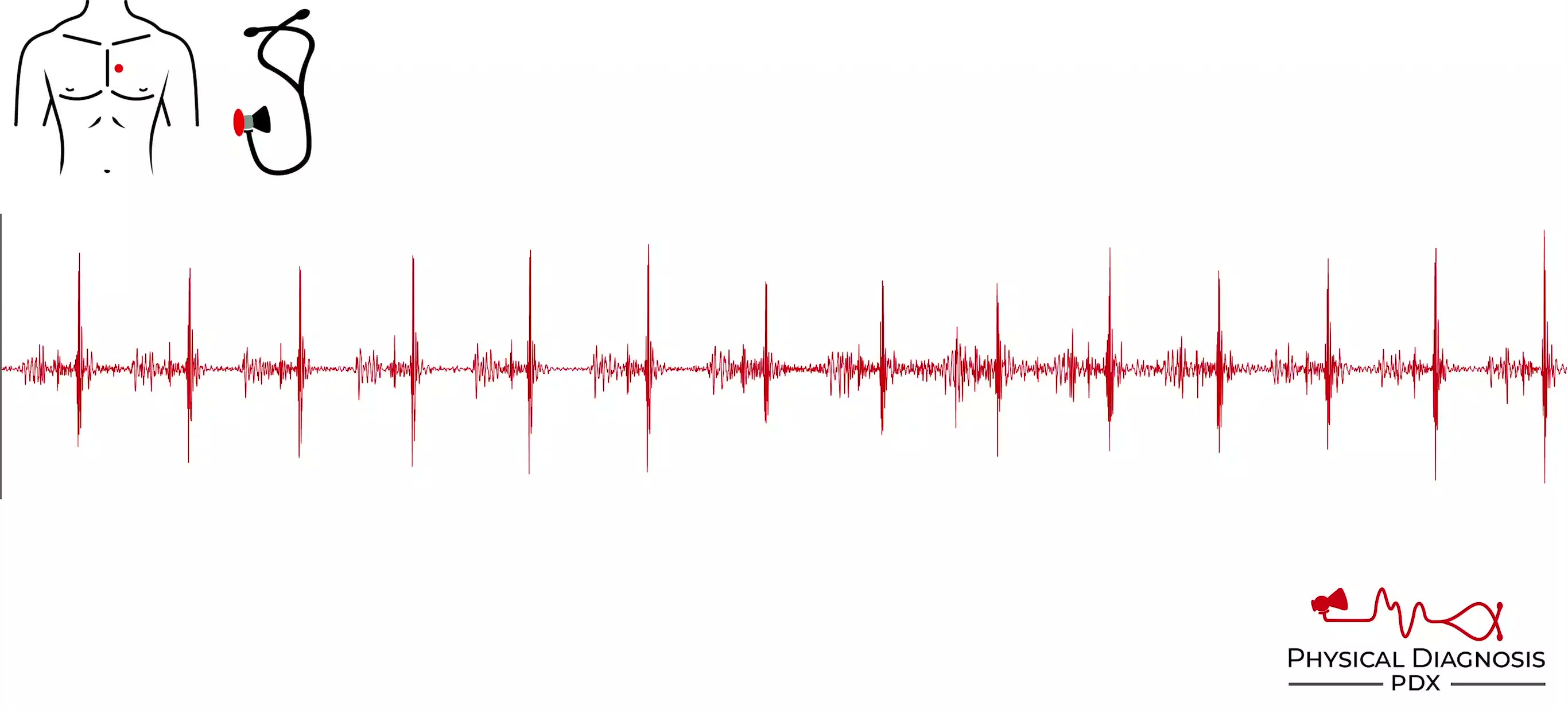
Tumor Plop
1 patients · 10 clips
The tumor plop is a high-pitched sound best heard with the diaphragm of the stethoscope. The classic tumor plop originates from an atrial tumor obstructing the mitral orifice, resulting in an early diastolic sound from e
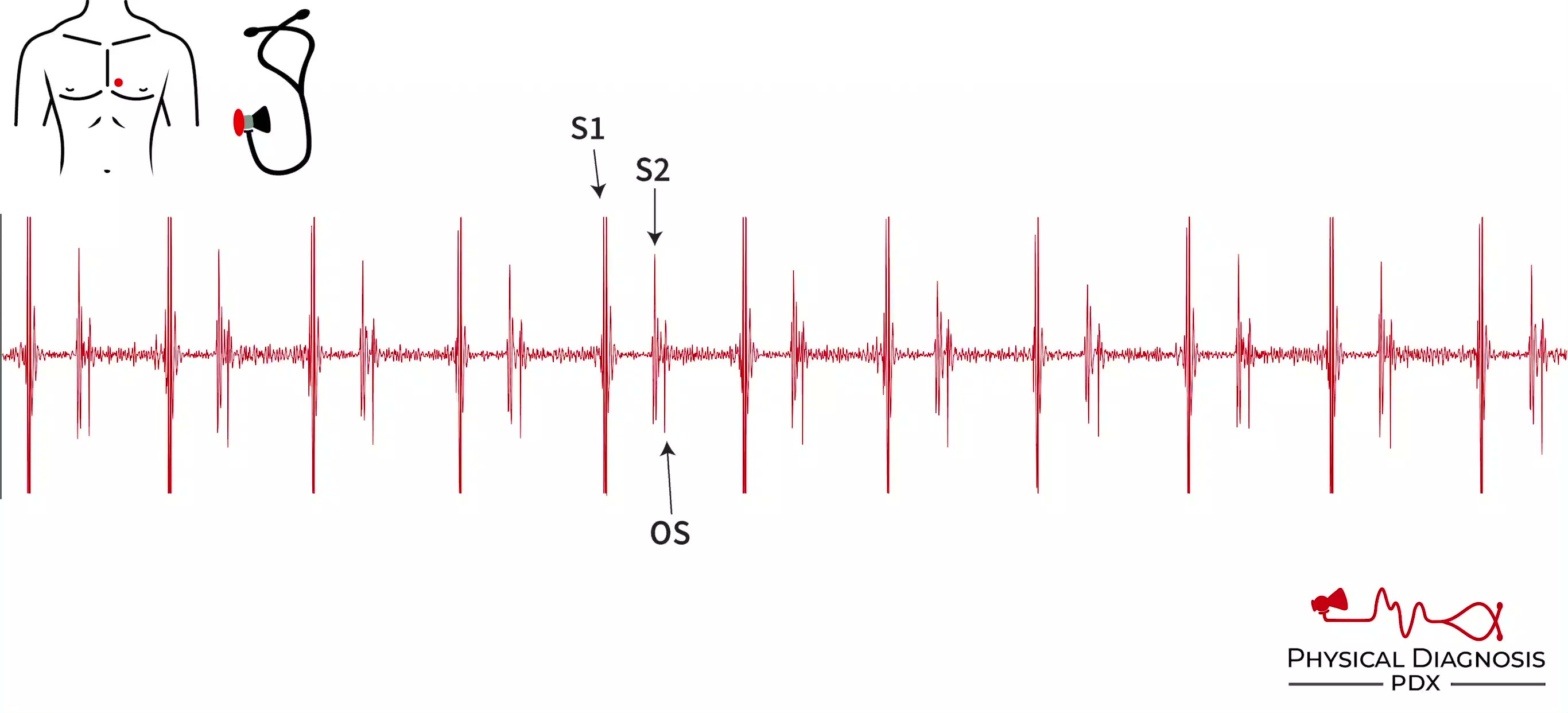
Opening Snap
2 patients · 4 clips
The opening snap is a high-pitched sound in early diastole that occurs in the vast majority of patients with mitral stenosis. It coincides with the opening of the mitral valve. It generally occurs closer to S2 than the p
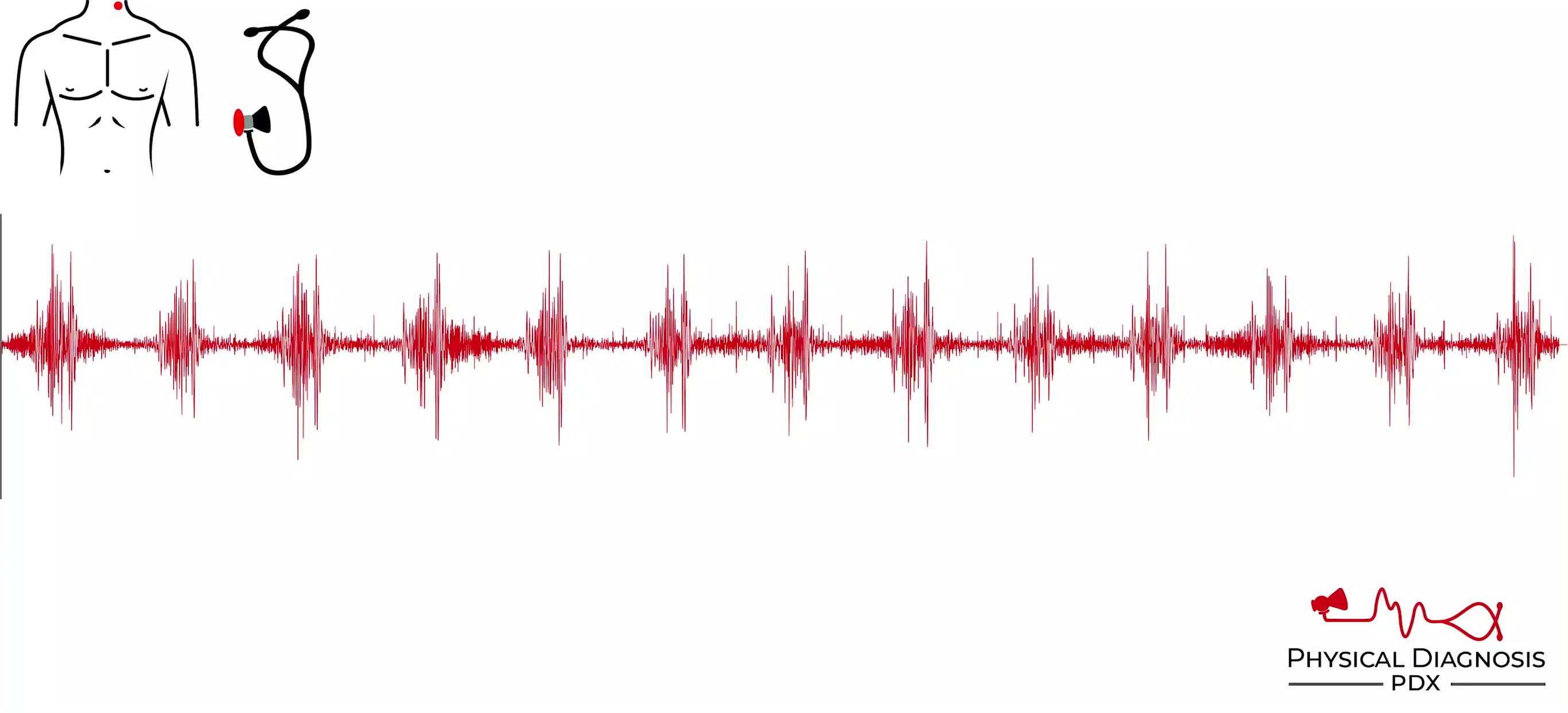
Aortic Coarctation
1 patients · 2 clips
Patient 1: This is a middle-age man who was admitted to the hospital with cardiogenic shock of unclear etiology. He was discovered to have Corrigan’s pulse on exam, which led to the discovery of Quincke’s pulse, radial-f
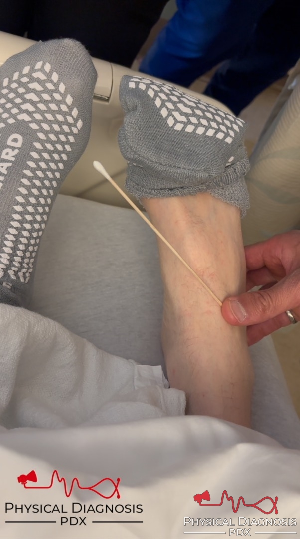
Bounding Pulse
6 patients · 11 clips
Large bounding pulses are seen in aortic insufficiency and other high-output states, such as thyrotoxicosis, severe anemia, right-to-left shunt, and wet beriberi. Card Tutorial Patient 1: This is a young man with intrave
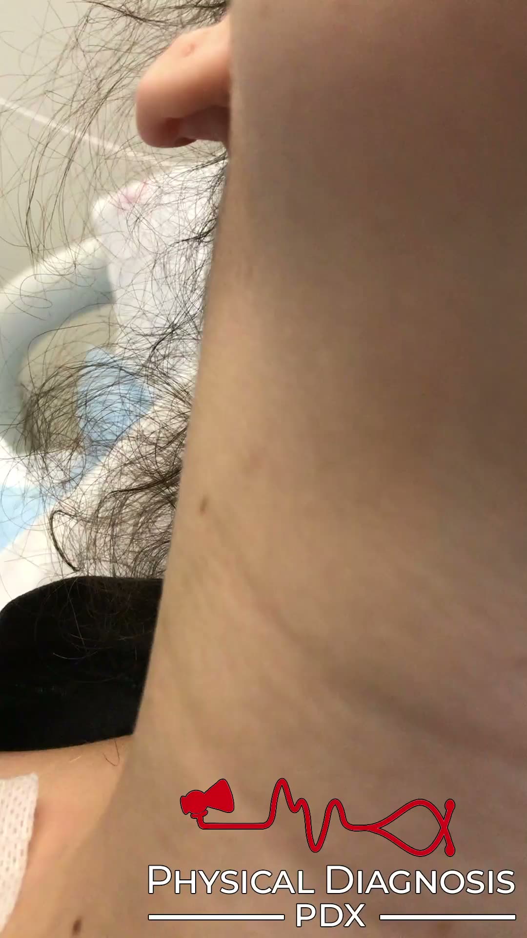
Carotid Pulse
5 patients · 7 clips
The jugular venous pulse must be distinguished from the carotid pulse. There are important differences between arterial and venous pulses that allows the examiner to distinguish them at the bedside. The normal carotid ar
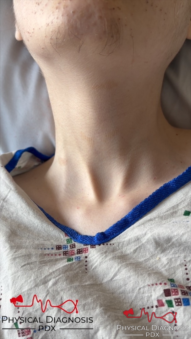
Corrigan’s Pulse
8 patients · 14 clips
A large, bounding carotid pulse seen in aortic regurgitation, high-output physiology (sepsis, liver disease, wet beriberi, etc.), and coarctation of aorta. Card Tutorial Patient 1: This is an older man with an aortic roo
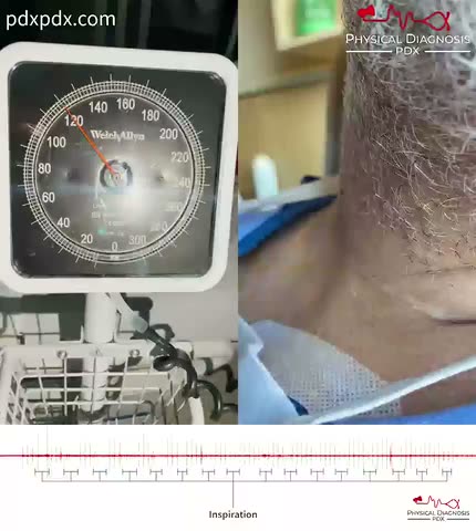
Pulsus Paradoxus
1 patients · 1 clips
A decrease in SBP >10 mmHg during inspiration. Consistent with cardiac tamponade or acute asthma attack. **Patient 1:** This is an older man with effusive-constrictive pericardial disease who developed presyncope and hyp
Pulsus bisferiens
1 patients · 1 clips
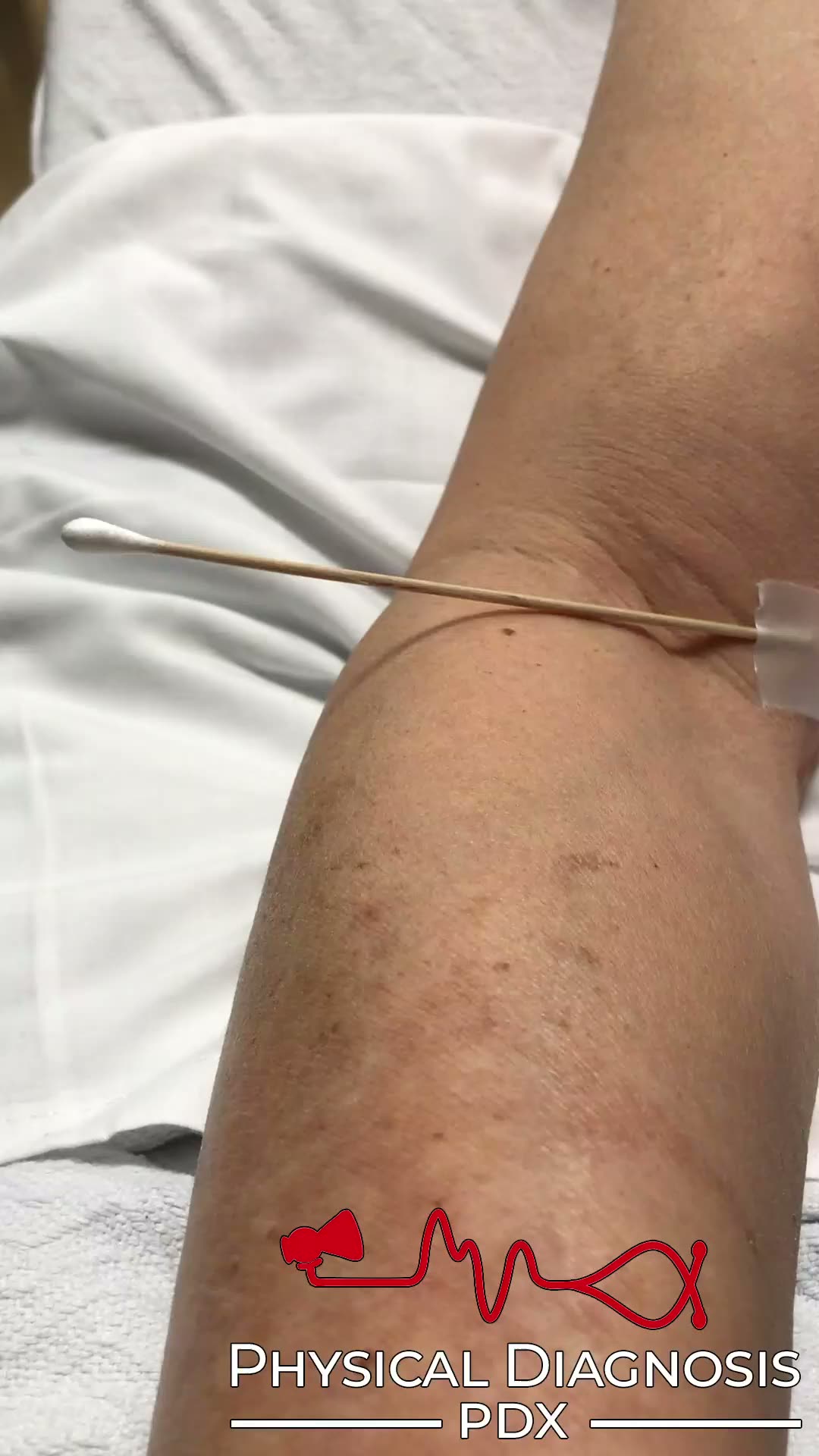
Pulsus bisferiens describes the presence of an exaggerated “double pulsation” in the arterial pulse. While the arterial pulse is normally dicrotic, consisting of a percussion wave and a tidal wave, the individual waves a
Radial- Femoral Pulse Delay
1 patients · 2 clips
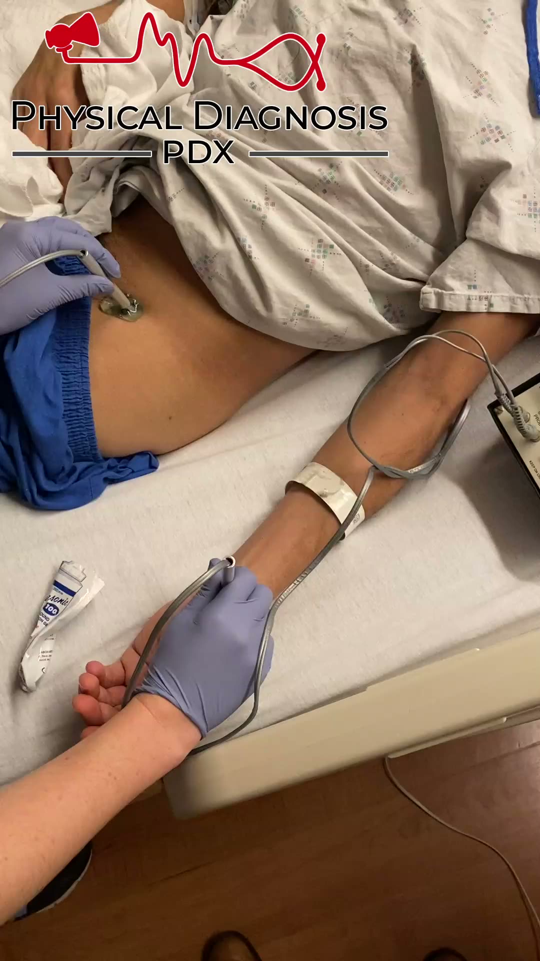
Patient 1: This is a middle-age man who was admitted to the hospital with cardiogenic shock of unclear etiology. He was discovered to have Corrigan’s pulse on exam, which led to the discovery of Quincke’s pulse, radial-f
Vigorous Carotid Pulse
1 patients · 2 clips
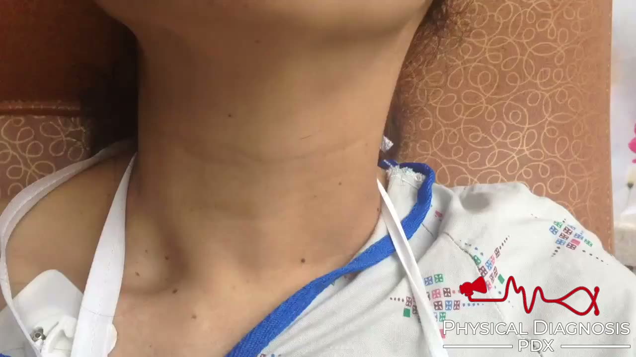
Coarctation of the aorta is a common congenital condition, accounting for 7% of all congenital heart lesions. An augmented carotid pulse – “carotid swell” – can be a clue to the diagnosis. A sustained systolic arterial w
Vigorous Peripheral Pulse
3 patients · 4 clips
Large bounding pulses are seen in aortic insufficiency and other high-output states, such as thyrotoxicosis, severe anemia, right-to-left shunt, and wet beriberi. Pulmonology Tutorial Patient 1: This is a middle-aged man